

Gastroenterology
Diagnostic and therapeutic endoscopy, oesophageal manometry and pH studies for reflux and swallowing problems, ERCP for the bile duct, FibroScan and hepatology, and long-term care for IBD, IBS and coeliac disease.
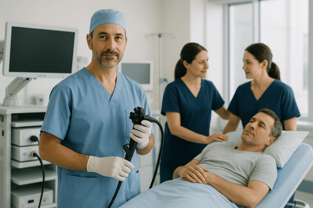
From the symptom you can live with to the finding you cannot ignore
Most people arrive with reflux that no longer answers to tablets, a bowel habit that has changed, or a liver result flagged on a routine blood test. The work is deciding which of those needs a camera this week and which needs a plan for the next year.
The bowel, IBD and IBS
Screening and polyp removal, the difference between an irritable bowel and an inflamed one, and the long-term care that follows a diagnosis.
Reflux and swallowing
Where the unit is strongest: measuring reflux properly before treating it, and the pressure studies that explain why swallowing has changed.
Liver, bile duct and pancreas
Fatty liver assessed without a biopsy, hepatitis treated, stones cleared from the bile duct, and the pancreas watched properly.
A hospital, not an endoscopy clinic
Endoscopy is easy to buy and hard to do well. The difference is not the camera: it is what happens when the camera finds something: a pathologist who reads the biopsy the same week, a surgeon down the corridor when a lesion is too large to remove endoscopically, and an oncology team already in the building if the result is the one nobody wanted.
It also means the honest answer is available. A unit that only performs procedures has an incentive to find a reason to perform one. A unit that has to live with the follow-up does not.
What we will not do
- Repeat an endoscopy that does not need repeating. Reports and slides travel; we ask for them first.
- Treat reflux surgically without measuring it. A procedure built on symptoms alone is a procedure built on a guess.
- Present an endoscopic weight procedure as equivalent to bariatric surgery.
- Quote a price for a procedure before anyone has decided that you need it.
- Promise a diagnosis in one visit when the honest answer depends on a biopsy that takes its own time.
Gastroenterologists who lead this work
What actually happens, in order
Send the report and the images
Endoscopy reports are far more useful with the photographs and the pathology than without them. Send the biopsy result itself, not only the letter that mentions it.
Consultant review
What the existing endoscopy already settles, and what genuinely needs repeating. Slides can often be re-read rather than the biopsy redone.
Tests on arrival
Endoscopy and colonoscopy on the same admission where that is safe, with manometry, pH studies and FibroScan arranged around them.
The procedure day
Most diagnostic endoscopy is day-case with sedation. ERCP, large resections and anything with a stent are planned as inpatient work.
Before you fly
Pathology explained in person where the timing allows, a written plan for the medicines you leave on, and a named route back for the results that arrive later.
Six things worth knowing first
Rectal bleeding is investigated
Assuming it is haemorrhoids is the commonest way bowel cancer is missed. Haemorrhoids are extremely common, which is exactly why they are a dangerous explanation to settle on.
Vomiting blood is an emergency
Blood, material that looks like coffee grounds, or black tarry stools all mean bleeding somewhere in the digestive tract. It is investigated as a hospital emergency, not as a clinic appointment.
Fever with jaundice is different
Yellow eyes and pain under the right ribs with fever and shivering can mean an infected, blocked bile duct. That combination needs hospital assessment immediately.
Test for coeliac before you stop gluten
Going gluten-free first makes the blood tests and the biopsy unreliable, and the only way back is to eat gluten again for weeks. Sequence matters here.
The prep is the procedure
A colonoscopy is only as good as the view. An incomplete preparation means polyps are missed and the whole thing is repeated, which is why the instructions are strict.
Endoscopic weight procedures are not surgery
A gastric balloon or an endoscopic sleeve is a different intervention with different limits. Anyone presenting them as equivalent to bariatric surgery is selling, not advising.
Jump to what you came for
Quick answer
Gastroenterology is the medical unit that diagnoses and treats disorders of the digestive system, including the esophagus, stomach, intestines, liver, pancreas, and gallbladder. At Acibadem in Turkey, gastroenterology care includes evaluation with laboratory tests, imaging, and endoscopic procedures, followed by personalized medical treatment and coordinated follow-up when advanced intervention or surgery is needed.
What this unit covers
Gastroenterology at Acıbadem International is built around the endoscopy suite and the clinics that feed it. Most people arrive with a symptom rather than a diagnosis: heartburn that no longer responds to tablets, food that sticks, a change in bowel habit, an abnormal liver blood test, a stool screening result that came back positive. The work is to find out what is actually happening, treat what can be treated without an operation, and be honest about the point where a different team should take over.
The scope of the unit is set out here so you can see whether your problem belongs with us. Each item has a section of its own that explains it properly.
Endoscopy and the tests that go with it
- Upper endoscopy, also called gastroscopy or EGD, for the oesophagus (esophagus), stomach and duodenum.
- Colonoscopy, including preparation, alternatives to colonoscopy, and everything about sedation, comfort and risk.
- Endoscopic ultrasound for staging and for sampling lesions in the pancreas and the wall of the gut.
- Capsule endoscopy and deep enteroscopy for the small bowel, the part no ordinary scope reaches.
Bowel cancer prevention
- Bowel cancer screening: who should be screened and when.
- Polyps and polypectomy, including removal at the same session.
- EMR and ESD for large lesions that would otherwise mean bowel surgery.
- The follow-up interval after polyps have been found, and what it depends on.
- Detection and staging of cancers of the stomach, oesophagus, pancreas and liver.
Reflux, swallowing and the muscles of the oesophagus
- Reflux and GERD, and where medication stops being the answer.
- pH and impedance monitoring, the tests that measure reflux rather than assume it.
- Oesophageal manometry, including high-resolution studies.
- Achalasia and the POEM procedure.
- Oesophageal dilation for strictures and rings.
- TIF and the assessment for fundoplication, including the role of a hiatal hernia.
- Barrett’s oesophagus, its surveillance and ablation.
- Difficulty swallowing, including eosinophilic oesophagitis.
Stomach, bowel and anorectal conditions
- Helicobacter pylori: testing, treatment and the retest that is often skipped.
- Gastritis and peptic ulcer.
- Irritable bowel syndrome and SIBO.
- Crohn’s disease and ulcerative colitis, from diagnosis to long-term monitoring.
- Coeliac (celiac) disease and food intolerance.
- Haemorrhoids, fissures and anorectal problems, including anorectal manometry.
- Bleeding and the symptoms that must not wait.
Liver, pancreas and bile ducts
- Fatty liver disease, now called MASLD.
- FibroScan and liver elastography for measuring scarring without a biopsy.
- Cirrhosis and portal hypertension, including varices surveillance.
- Hepatitis B and hepatitis C.
- Gallstones and bile duct stones.
- ERCP for stones and blocked ducts.
- Pancreatitis and pancreatic cysts.
- Endoscopic weight procedures, and nutrition support including feeding tubes.
What this unit does not do itself
Cancer treatment is not run from here. We detect, sample and stage; chemotherapy, radiotherapy and the tumour board belong to Medical Oncology, and we hand over at that point rather than pretending otherwise. Liver transplantation belongs to the Organ Transplantation unit; we assess and manage cirrhosis and its complications and hand over when transplant becomes the question.
Operations are not ours either. Gallbladder removal, bowel resection, hernia repair and haemorrhoid surgery belong to General Surgery, and weight-loss surgery to Bariatric Surgery. What we own is the endoscopic and medical side, and the judgement about when an operation is genuinely the better answer. If your problem needs a surgeon, we will say so plainly instead of offering an endoscopic version of an operation that will not hold.
Upper endoscopy (gastroscopy, EGD)
An upper endoscopy is a direct look at the lining of the upper digestive tract with a thin flexible camera passed through the mouth. It is called gastroscopy in most of Europe and EGD, short for oesophagogastroduodenoscopy, in most of the United States. The test and the instrument are the same. It is the single most useful test in gastroenterology because it does three jobs at once: it looks, it takes tissue, and it can treat.
What a gastroscopy can see
The scope passes down the oesophagus (esophagus), into the stomach, through the pylorus and into the first and second parts of the duodenum. Along the way the endoscopist is looking for inflammation, ulcers, narrowings, hernias, varices, changes in the colour and pattern of the lining, and anything raised, flat or ulcerated that should not be there.
Modern scopes give a magnified, high-definition image, and light filters can be switched on that make the surface blood vessels and pit patterns stand out. This matters because the most important early changes in the stomach and oesophagus are flat and subtle, not lumps. A careful, unhurried examination with good views of every wall finds things that a rushed one misses, which is why the endoscopist will inflate the stomach with air or carbon dioxide, wash the lining, and take their time in the areas that hide lesions.
What it cannot see is anything outside the wall or beyond the reach of the scope. The pancreas, the bile ducts and the deeper layers are assessed with endoscopic ultrasound, which is covered in its own section. The small bowel beyond the duodenum is reached with capsule endoscopy or deep enteroscopy.
The symptoms that make an upper endoscopy the right first test
Not every case of indigestion needs a camera. What moves a symptom from “treat and see” to “look now” is the presence of features that suggest something structural.
- Difficulty swallowing, or food that sticks and has to be washed down. This is never a symptom to watch and wait on.
- Pain on swallowing, or the sensation that swallowing has changed over weeks.
- Vomiting that persists, particularly vomiting of food eaten hours before.
- Unintended weight loss with upper abdominal symptoms.
- Iron deficiency anaemia without an obvious cause. The upper tract is one of two places to look, the colon being the other.
- Reflux that does not settle on proper treatment, or that comes straight back every time treatment stops.
- New indigestion in an older adult, or in anyone with a family history of stomach or oesophageal cancer.
- Suspected bleeding, including black stools or anaemia with no other source.
- A positive coeliac blood test, where duodenal biopsies confirm the diagnosis.
- Known cirrhosis, where the scope is used to look for varices; the surveillance logic belongs to Cirrhosis and portal hypertension.
How the test is done, from arrival to recovery
You will be asked not to eat for several hours so the stomach is empty; clear fluids are usually allowed until a couple of hours before, and the exact clock times come from the letter the unit sends you, which overrides any general rule. An empty stomach is not a formality. Retained food hides lesions, wastes the appointment and raises the risk of material going into the lungs while you are sedated.
From the door to the recovery bay, the sequence runs like this.
- You are checked in, your medicines and allergies are reviewed, and the consent conversation happens while you are still fully alert.
- A cannula goes into a vein in the back of your hand if you are having sedation.
- Your throat may be sprayed with a local anaesthetic, which tastes bitter and makes the throat feel swollen, though nothing is actually swollen.
- A small plastic mouthguard is placed between your teeth to protect them and the scope.
- You lie on your left side, and the scope is passed while you swallow once. After that you do not need to swallow again.
- Air or carbon dioxide is used to open the folds so the walls can be inspected, which is what causes the bloated feeling and the burping. Carbon dioxide is absorbed quickly and leaves less wind afterwards than air.
- You rest in recovery until the sedation and the throat spray have worn off enough to swallow safely.
The examination itself is short, usually shorter than the checking-in and the recovery that surround it. You can breathe normally the whole time, because the scope goes into the oesophagus, not the airway. This is the single most common fear and it is worth stating plainly: the tube does not block your breathing.
Afterwards you rest until the sedation wears off and the throat spray has worn off enough to swallow safely. The choice of sedation, the monitoring, the escort rule and the rules about driving are all set out in Sedation, comfort and risks, which owns that ground for every procedure in this unit.
Ultrathin and transnasal endoscopy
Some units offer a much thinner scope passed through the nose rather than the mouth. It avoids the gag reflex almost entirely and many people tolerate it with no sedation at all, which means no cannula, no recovery period and no escort home. It is a reasonable option for a straightforward diagnostic look, particularly if you have had a bad experience with a standard gastroscopy, or if avoiding sedation matters for medical reasons.
The trade-off is honest: a thinner scope has a smaller working channel, so taking multiple biopsies is slower and most therapeutic work cannot be done through it. If the plan includes dilation, treating a bleeding point or removing something, the standard scope is the right instrument. Whether an unsedated route suits you is a decision taken with the endoscopist, and the sedation options themselves are described in Sedation, comfort and risks.
Why biopsies are taken even when the lining looks normal
Biopsies are small pinches of tissue taken with forceps passed down the scope. You do not feel them. The lining of the gut has no fine touch sensation of the kind your skin has, so people are routinely surprised to be told afterwards how many samples were taken.
They are taken from normal-looking tissue for a reason: several of the most important diagnoses in the upper gut are invisible to the eye and only exist under a microscope.
- Helicobacter pylori. The stomach lining can look near enough normal while the bacterium is present. Testing and treatment are covered in Helicobacter pylori, which owns all of that ground including the breath test and the retest.
- Coeliac disease. The duodenum often looks unremarkable, and the diagnosis is made on the biopsy. One sequencing point matters enough to repeat here: do not start a gluten-free diet before the test, because it can turn a positive result negative. The full explanation is in Coeliac disease and food intolerance.
- Eosinophilic oesophagitis. Diagnosed by counting cells in oesophageal biopsies, and sometimes the only abnormality in a person whose food keeps sticking. See Difficulty swallowing.
- Barrett’s oesophagus. Suspected by the appearance, confirmed and graded on biopsy. See Barrett’s oesophagus.
- Atrophy and intestinal metaplasia of the stomach lining. These change how closely the stomach needs watching in future.
Biopsies take days to process, so a normal-looking examination does not mean the answer is complete on the day. If you are travelling, ask before you leave how the histology result will reach you.
Treatments carried out during the same examination
An upper endoscopy is often a treatment rather than just a test. What can be done through the scope in the same session includes stopping a bleeding ulcer with clips, injection or heat; removing food that has become stuck; banding oesophageal varices; stretching a narrowing; removing polyps of the stomach or duodenum; and placing a feeding tube through the abdominal wall.
Each of those has its own home. Dilation is described in Oesophageal dilation, variceal work in Cirrhosis and portal hypertension, feeding tubes in Feeding tubes and nutrition support. The point for a patient making a plan is simply this: if a treatable problem is found, you may wake up having had it treated, and the consent conversation before the procedure will cover that possibility.
What a normal gastroscopy does and does not rule out
A normal result is genuinely reassuring, and it is a real answer rather than an absence of one. But it is reassuring about a defined list, and that list has an edge.
| Ruled out by a normal gastroscopy | Not ruled out by it |
|---|---|
| Ulcers | The pancreas |
| Tumours of the lining | The gallbladder and the bile ducts |
| Coeliac disease | The colon |
| Strictures | The small bowel past the duodenum |
| Inflammation of the upper tract | Reflux |
Reflux is the entry on that list that surprises people most. Many people with genuine, damaging reflux have a normal-looking oesophagus, which is exactly why reflux is measured rather than assumed, as described in Measuring reflux: pH and impedance monitoring.
And a normal gastroscopy in someone with persistent symptoms often points towards a functional disorder: a gut that is working abnormally rather than one that is structurally damaged. That is a diagnosis, not a dismissal, and it has treatments of its own.
Your report
You should leave with a written report describing what was seen, where any biopsies were taken from and what happens next. Ask for images if you want them; most units can print or send them. If you have travelled, ask for the report in English and check that the histology pathway is written down.
Recovery is usually uneventful. A sore throat for a day, wind and mild bloating are expected. What is not expected is pain.
Colonoscopy
A colonoscopy is an examination of the whole large bowel with a flexible camera passed through the anus. It is the only test that both finds and removes precancerous polyps in the same sitting, which is why it sits at the centre of bowel cancer prevention rather than merely at the centre of diagnosis.
What the examination covers
The scope is advanced around the colon to the caecum, where the small bowel joins, and often a short distance into the last part of the small bowel. The examination that counts is the one performed on the way out: the endoscopist withdraws slowly, washing, insufflating and flattening the folds so that the flat lesions hiding on their far side are seen.
A flexible sigmoidoscopy is the same instrument used for a shorter look, reaching only the left side of the colon. It needs less preparation and no sedation in most people, and it is useful for assessing rectal bleeding or the extent of colitis. It is not a substitute for a full colonoscopy when the whole bowel needs to be seen, because a right-sided lesion will simply not be reached.
Colonoscopy age: when a first colonoscopy is usually offered
For people at average risk with no symptoms, several major guidelines moved the starting age for bowel cancer screening from 50 to 45, and national programmes have not all followed at the same pace; some still begin at 50 and some screen with stool tests first, sending only positive results for colonoscopy. So the honest answer to “what age should I have a colonoscopy?” is that the colonoscopy age depends on which country’s programme applies to you and on your personal risk, not on a single universal number.
How family history changes that start date, and how long screening should continue into later life, are set out in Bowel cancer screening.
Reasons to have a colonoscopy sooner than the screening age
Screening ages describe people without symptoms. They do not apply to someone with a symptom, and this is the most common and most dangerous misunderstanding we see. A thirty-year-old with rectal bleeding does not “wait until 45”; they get assessed.
- Bleeding from the rectum, or blood mixed into the stool.
- A persistent change in bowel habit lasting weeks, particularly looser stools or a new need to go urgently.
- Iron deficiency anaemia without an explanation.
- Unintended weight loss with bowel symptoms.
- Ongoing abdominal pain with a change in bowel pattern.
- A positive stool screening test of any kind.
- A first-degree relative with bowel cancer or advanced polyps, or a known inherited syndrome in the family.
- Long-standing ulcerative colitis or Crohn’s colitis, where surveillance follows a different schedule; see Crohn’s disease and ulcerative colitis.
What the examination itself is like
You change into a gown, a cannula is placed if you are having sedation, and you lie on your left side with your knees drawn up. Most people in this unit have sedation, and the options and their trade-offs are set out in Sedation, comfort and risks.
Carbon dioxide is used to open the bowel so the walls can be inspected. It is absorbed through the bowel wall and breathed out, so the bloating settles quickly compared with room air. During the examination you may be asked to change position or a nurse may press on your abdomen; both are ordinary manoeuvres to help the scope round a bend rather than signs that something is wrong.
Discomfort, when it happens, is a stretching or cramping sensation as a loop of bowel is straightened, and it comes in waves rather than continuously. If a polyp is found it is usually removed there and then, which you will not feel. What polyps are and how they are removed is covered in Polyps and polypectomy, and larger lesions in EMR and ESD: removing large lesions without surgery.
Why the quality of the view decides the value of the test
A colonoscopy is only as good as the bowel it looks at. Residual stool hides flat lesions, and a poorly prepared colon produces a result that reads as normal but is not reliable: the worst possible outcome, because it gives false reassurance and a long interval before anyone looks again.
This is why units grade the quality of preparation in the report and will offer a repeat, sometimes soon, when the view was inadequate. It is also why the preparation instructions are specific and unforgiving about timing. The entire preparation schedule, what to eat on which day, when to take each dose and which medicines to hold, belongs to Preparing for a colonoscopy and is not restated anywhere else, so that there is only ever one set of timings to follow.
Reading your colonoscopy report
A good report tells you four things: how far the scope reached and whether the caecum was seen, how clean the bowel was, what was found and where, and what was removed or biopsied. If any of those four is missing, ask.
If polyps were removed, the tissue goes to the laboratory and the microscopic result is what determines when you should be examined again. That interval is decided by what was found and how completely it was removed, and the rule is explained in What happens after polyps are found. Nobody should leave a colonoscopy with a date for the next one before the histology is back, unless the finding was clear-cut enough to make the plan obvious on the day.
The hours after a colonoscopy
Expect wind, some cramping until the gas has passed, and possibly a small amount of blood on the paper if a polyp was removed or a biopsy taken. Eating starts with something light and moves back to normal the same day unless you are told otherwise. If you had polyps removed, you may be asked to avoid heavy lifting and long flights for a short period; the instruction is individual, so follow the one you are given.
Colonoscopy preparation: the prep, step by step
This section owns the whole colonoscopy preparation schedule: the days beforehand, the colonoscopy prep diet, the medicines to hold, the timing of each dose and the morning of the procedure. No other section restates these timings, so that you are never following two versions of the same instruction.
One rule sits over everything written here. Your unit’s own instruction sheet, with its clock times and its named product, is the one you follow. Preparations differ in volume, taste and dosing, and the letter you were sent is written for the product you were given and the time of your appointment. What follows explains the shape of the process, why each step exists and where people go wrong, not a substitute for that letter.
Why the colonoscopy prep is the hard part, and why it is not optional
Almost everyone who has had a colonoscopy says the same thing: the procedure was nothing, the preparation was the ordeal. That is an honest description and there is no point pretending otherwise. You will spend an evening and part of a night close to a toilet, drinking a large volume of liquid you would rather not drink.
It is worth doing properly because a bad preparation wastes the whole exercise. Stool left behind covers exactly the flat, subtle lesions the test exists to find, and the result is either a repeat within a short interval or, worse, a normal report that was never trustworthy. People who cut the prep short are not saving themselves a bad night; they are usually buying themselves a second one.
The colonoscopy prep diet: the days before, eating for a clear view
The aim is to reduce the residue moving through the bowel before the purge begins, so the laxative has less work to do.
- About five days before, stop foods that leave hard fragments behind: seeds, nuts, popcorn, sweetcorn, dried fruit, tomato and pepper skins, and any seeded bread. These are the particles that persist and sit behind folds.
- Two to three days before, move to a low-fibre diet. White bread, white rice and pasta, eggs, plain chicken or fish, potato without skin, clear soups, cheese, yoghurt without fruit pieces. No wholegrains, no raw vegetables, no salad, no fruit skins, no pulses.
- The day before, most units ask for clear fluids only from a set time, often from breakfast or lunchtime. Some allow a light low-fibre breakfast first. Follow your letter on this point in particular, because it varies more than any other step.
- Drink more than you think you need throughout the day before. Dehydration is the main cause of feeling dreadful during the prep, and the extra clear fluid is part of the preparation rather than an optional extra: this matters most with the low-volume products, where people assume the small bottle is the whole requirement.
Medicines that need a decision before the prep starts
Raise these at the time of booking, not the night before. Some need a decision from the doctor who prescribed them, and that takes days to arrange.
- Blood thinners and antiplatelet drugs. Never stop these on your own initiative. Whether they are held, and for how long, depends on which drug it is, your kidney function, why you take it: a mechanical heart valve, a recent stent and atrial fibrillation are not the same situation, and how likely polyp removal is. The decision belongs to the prescribing doctor together with the endoscopist. Aspirin taken long term is often continued.
- Iron tablets. Stop about five days before, or as your unit instructs. Iron turns the stool black and sticky and coats the lining, and it is one of the commonest reasons a preparation looks poor despite the patient having done everything else correctly.
- Diabetes medication. A day of clear fluids changes everything about your glucose control. Insulin doses usually need adjusting, some tablets are held, and drugs of the SGLT2 inhibitor class are generally paused around the procedure. Ask your diabetes team for a written plan and take your meter with you.
- GLP-1 weight and diabetes injections. Tell the team you are taking one. These drugs slow stomach emptying, guidance about them is still changing, and you may be asked to pause the injection before a procedure involving sedation.
- Constipating medicines. Opioid painkillers, some antidepressants and anti-diarrhoeal drugs make the bowel harder to clear and may need an extended preparation.
- Bulk-forming fibre supplements. Stopped several days before, along with the high-residue foods.
- Everything else, including blood pressure tablets, thyroid medication, epilepsy medication, immunosuppressants and inhalers, is usually continued. Do not stop a regular medicine because it is not on a list; ask instead.
The purge itself: why it is split into two doses
The laxative is taken in two parts rather than all at once, and this is not a matter of comfort. The colon keeps producing mucus and residue overnight, so a preparation finished entirely the evening before leaves the right side of the bowel and the caecum coated by morning. Splitting the dose, with the second half taken on the day of the procedure, is what produces a clean view of the very part of the colon where lesions are easiest to miss.
- First dose: the evening before, at the time your letter gives. Expect it to start working within an hour or two, though this varies widely between people.
- Second dose: in the early hours or the morning of the procedure, at the time your letter gives, typically some hours before the appointment and finished a set interval before it. That final gap exists so the stomach is empty for sedation. It is a safety interval set by your unit, not a suggestion, and the letter’s times are the ones to follow.
- An afternoon appointment shifts everything later rather than removing the second dose. Do not decide on your own to take both doses the night before to get a better night’s sleep: it is the single most effective way to ruin the test.
If your appointment is very early and the timings in your letter look impossible, telephone the unit rather than improvising. The times can usually be adjusted; a preparation you invented cannot be corrected.
Getting the liquid down without being sick
The volume defeats more people than the taste does. A few practical things genuinely help.
- Chill the made-up solution in the fridge. Cold blunts the taste considerably.
- Drink it through a straw placed towards the back of the tongue, which bypasses much of the taste.
- Pace it: a glass every ten to fifteen minutes rather than heroic gulps. Rushing causes nausea and vomiting, and a vomited dose is a lost dose.
- Take a sip of a permitted clear drink between glasses to clear the taste, apple juice, clear lemon squash or ginger ale, none of them red or purple.
- Suck a clear boiled sweet if allowed, and keep barrier cream and moist wipes by the toilet. Repeated wiping is what makes the night sore, not the laxative itself.
- If you vomit a dose, stop for half an hour, then restart slowly rather than abandoning it. If you cannot keep it down at all, telephone the unit.
What counts as a clear fluid
The rule is simple: if you can see through it and it is not red or purple, it is usually allowed. Red and purple dyes stain the lining and can be mistaken for blood.
| Allowed | Not allowed |
|---|---|
| Water, still or sparkling | Milk, cream, and anything containing them |
| Clear apple or white grape juice | Orange juice, smoothies, juice with pulp |
| Black tea or black coffee, no milk | Coffee with milk, milky tea, hot chocolate |
| Clear broth or consommé | Soup with pieces, creamed soup |
| Clear fizzy drinks, sports drinks | Anything red, purple or blackcurrant |
| Clear jelly, honey, clear boiled sweets | Alcohol, and all solid food of any kind |
Sugary clear fluids are useful rather than forbidden during the prep day: they provide something to run on when you are not eating. If you have diabetes, sugar-free versions and your team’s plan take priority.
How to tell the preparation is working
What you are aiming for is stool that becomes progressively looser and paler until what you are passing is a clear or pale yellow liquid with nothing solid in it. Yellow is normal and expected; it is bile, not a problem. Small flecks may persist and that is usually acceptable.
What is not acceptable is still passing brown, cloudy or formed stool when you have finished the whole preparation. Telephone the unit rather than turning up hoping for the best. They can advise an extra dose, a supplementary laxative or, occasionally, rescheduling, and they would far rather hear from you at six in the morning than write “inadequate preparation” in your report.
Feeling cold, shivery, light-headed or having a headache usually means you have not drunk enough clear fluid alongside the preparation. Drink more, and telephone the unit if it does not improve.
The morning of the procedure
- Take the second dose at the time given, and finish it by the deadline in your letter.
- Stop all clear fluids at the time you were told, generally two hours before your appointment. After that, nothing by mouth.
- Take the morning medicines you were told to continue with a small sip of water.
- Leave jewellery and valuables at home, wear something loose, and bring your medication list, your allergy list and any previous endoscopy or pathology reports, particularly if you are being seen in a country where your records are not already in the system.
- Bring the responsible adult who will take you home. If you are having sedation, this is not negotiable and the arrangements are explained in Sedation, comfort and risks.
- Expect to be in the department for several hours in total. The procedure is the short part of the visit.
Preparation when you have diabetes, kidney or heart disease
Bowel preparation is a genuine physiological event, not simply an inconvenience. It shifts fluid and salts, and in some people that matters.
If you have kidney disease, heart failure, liver disease with fluid retention, or you take diuretics or drugs that affect kidney function, tell the unit before the prep is chosen. Certain preparations are avoided altogether in reduced kidney function, and others need a specific plan for how much you drink and when your regular medicines are taken. Never buy a bowel preparation online or use one left over from a relative’s appointment: the choice of product is a medical decision made on the basis of your kidneys, your heart and your medication list.
Older and frailer patients, and anyone who lives alone, should say so. Preparations can be spread over a longer period, and having someone in the house overnight is a reasonable request rather than fussiness.
When the standard colonoscopy prep is not enough
Some people need more. Long-standing constipation, opioid use, previous abdominal surgery with adhesions, diabetes of long duration, and any history of a poor preparation all predict a bowel that clears slowly. In those situations the answer is a longer preparation, starting the low-fibre diet sooner and adding a laxative for several days beforehand, rather than the same regimen taken more hopefully.
Tell the unit if a previous colonoscopy was abandoned or reported as poorly prepared. That single piece of history changes the plan and is very often the difference between a conclusive examination and a repeated one. If you have a stoma, a known stricture, or you are on dialysis, the preparation is individualised and needs to be discussed well before the appointment.
Colonoscopy alternatives: when colonoscopy is not the only option
Colonoscopy is the reference test, but it is not the only way into bowel cancer screening, and for some people it is not the right first step. The colonoscopy alternatives are legitimate tests, not second-best ones. What they share is one structural limitation that must be understood before choosing any of them: none of them can remove a polyp. Every one of them, when abnormal, ends in a colonoscopy.
Faecal immunochemical testing (FIT)
FIT looks for human haemoglobin in a small stool sample collected at home with a stick and a tube. It has replaced the older guaiac occult blood test in most programmes because it detects human blood specifically, needs only one sample in most schemes, and does not require any dietary restriction beforehand.
It is the backbone of national screening programmes for a reason: it is cheap, needs no preparation, no sedation and no time off, and it is repeated at intervals so that a bleed missed on one round is likely to be caught on the next. Its limitation is that it detects bleeding, and polyps often do not bleed. A normal FIT is reassurance about the interval, not a certificate that the colon is clear.
Multi-target stool DNA testing
Stool DNA tests look for altered DNA shed by abnormal cells as well as for blood. Combining the two makes them better than FIT alone at picking up advanced precancerous polyps, and they are done at home from a whole stool sample posted to a laboratory.
Two honest caveats. They produce more false positives than FIT, meaning more people are sent for a colonoscopy that turns out to be normal, and they are repeated at longer intervals, so a single test carries more weight. Availability also differs sharply between countries; it is not offered everywhere, and where it is not, FIT is the sensible stool test.
What a positive stool test means next
A positive stool test of any kind is not a diagnosis of cancer, and most people with one do not have cancer. It is a signal that the colon must be looked at directly, and the only test that can both look and remove what it finds is a colonoscopy.
This is the point where alternatives stop being alternatives. Repeating the stool test is not a substitute, and a normal repeat does not cancel the first result.
CT colonography, also called virtual colonoscopy
CT colonography is a CT scan of the abdomen with the colon gently distended with gas through a small rectal tube, reconstructed to give a view of the inside surface. There is no sedation, no scope and no recovery period, and it is completed in minutes.
It is a genuinely good option for people in whom a colonoscopy is unsafe or was incomplete: a scope that could not pass a narrowing, or a patient whose heart or lung disease makes sedation risky. It is also the standard way to see the colon beyond an obstructing tumour.
What it does not avoid is the preparation. The bowel still has to be clean, and most protocols add a contrast agent taken by mouth on the preparation days. Beyond that: it uses ionising radiation; it is less reliable for flat and small lesions; it cannot take a biopsy or remove anything, so a positive scan means a colonoscopy afterwards, with a second preparation; and it sees the rest of the abdomen too, which sometimes finds something incidental that leads to further tests of its own.
Colon capsule endoscopy
A colon capsule is a swallowed camera the size of a large tablet that photographs the bowel as it passes through, transmitting to a recorder worn on a belt. Nothing is passed into you, and there is no sedation.
The honest description is that it exchanges one difficulty for another. The preparation is heavier than for a colonoscopy, not lighter, because the images have to be clear from the inside of a moving capsule and additional laxatives are given during the test to move it along. It cannot take a biopsy or remove a polyp, so an abnormal result again ends in a colonoscopy. It is not suitable where there is a suspected narrowing, because a capsule can lodge in it. Small-bowel capsule endoscopy, which is a different indication altogether, is covered in Capsule endoscopy and deep enteroscopy.
Blood-based screening tests
Blood tests that look for tumour DNA circulating in the bloodstream have begun to appear for bowel cancer screening. The attraction is obvious: a blood sample takes minutes and people actually complete it.
The limitation is important and is often lost in the marketing. These tests are considerably better at detecting an established cancer than at detecting the advanced polyps that screening exists to remove before they become cancer. A test that finds cancer rather than preventing it is a weaker form of screening, even when it is more convenient. If you are choosing, understand which of the two things you are buying.
Choosing between the options honestly
The comparison that matters is not which test is theoretically best, but which one you will actually complete, and what you intend to do with an abnormal result.
- A stool test done reliably at every interval prevents more cancers than a colonoscopy that is endlessly postponed.
- Every non-colonoscopy option is a triage test. Choosing one means accepting in advance that an abnormal result commits you to a colonoscopy, with its preparation, after all.
- If you already have a reason to expect polyps, a family history, a previous polyp, long-standing inflammatory bowel disease, the triage tests are a poor fit, because the plan was always going to involve looking directly.
- If sedation or the scope itself is the barrier, say so rather than declining screening altogether. CT colonography, an unsedated examination or a different sedation plan may solve the actual problem; the options are set out in Sedation, comfort and risks.
When an alternative is the wrong answer
Screening tests are designed for people without symptoms. If you have symptoms, none of the options in this section is appropriate, because a normal result will not make the symptom safe: it will only delay the examination that could explain it. Rectal bleeding, a persistent change in bowel habit, unexplained iron deficiency anaemia or weight loss need a diagnostic examination, not a screening test.
Sedation, comfort and risks
This section covers sedation and complications for every endoscopic procedure in this unit, upper endoscopy, colonoscopy, and the therapeutic procedures built on them. It is written to be read before you consent, not afterwards.
Two things are true at once and both need saying. Endoscopy is among the safest invasive procedures in medicine, and serious complications are uncommon. And it is still an invasive procedure with real risks, which is why consent is taken, why you are monitored, and why the instructions afterwards are specific.
The sedation options and what each one feels like
- No sedation, with throat spray only (upper endoscopy). A local anaesthetic numbs the throat. You are fully awake and can drive yourself home. Some people find it entirely tolerable, particularly with an ultrathin scope; others find the gagging unpleasant.
- Conscious sedation. A sedative, usually with a short-acting painkiller, given through a cannula. You stay awake enough to follow instructions but relaxed and detached, and most people remember little or nothing afterwards. The amnesia is a normal effect of the drug rather than a sign you were unconscious.
- Deep sedation. A different agent, given and monitored by an anaesthetist (anesthesiologist), which puts you into a sleep you will not remember at all. It wears off quickly and cleanly, which is why it suits longer or more demanding procedures. It needs a person whose only job is your airway and your monitoring.
- General anaesthesia. Reserved for particular situations: some complex therapeutic procedures, a difficult airway, a high risk of material entering the lungs, or young children.
Sedation is not the same as general anaesthesia, and the difference is worth understanding: with sedation you are breathing for yourself throughout, and you may respond to voice or touch even though you will not recall it.
How the level of sedation is decided
It is a joint decision, and it is individual. What goes into it: which procedure is planned and how long it is likely to take; your age, weight, heart and lung health; whether you have obstructive sleep apnoea or a difficult airway; what happened at previous endoscopies; the medicines you take; and how anxious the prospect makes you, which is a legitimate clinical factor rather than a weakness.
Country of origin matters more than people expect. Unsedated gastroscopy is routine in some health systems and almost unheard of in others, so patients arriving from abroad often expect either much more or much less than is standard here. Say what you expect at the pre-procedure conversation rather than discovering the difference on the trolley.
Monitoring while you are asleep or drowsy
Throughout the procedure a nurse or anaesthetist watches your oxygen level, pulse, blood pressure and breathing continuously, and oxygen is given through the nose as a routine rather than as a sign of trouble. Sedation is reversible: agents exist that reverse the common sedative and painkiller if breathing becomes too shallow, and resuscitation equipment is in the room whether or not it is ever needed.
Your consent should be taken before any sedative is given. If you are asked to sign something once the cannula is in and the drug is running, that is the wrong order, say so.
Waking up, the escort rule and the rest of the day
You recover in the department until you are awake, your observations are stable and you can drink. Someone will tell you what was found before you leave, but sedation blurs memory, so ask for it in writing and, if you can, have your companion hear it too.
If you have had any sedation, for the rest of that day you must not drive, operate machinery, drink alcohol, sign legal or financial documents, or be solely responsible for a small child. A responsible adult must take you home and, ideally, stay with you overnight. This is not caution for its own sake: judgement and memory are impaired for hours after you feel normal. A taxi alone does not satisfy the rule in most units, and if you cannot arrange an escort, tell the unit in advance so the procedure can be planned unsedated instead of cancelled on the day.
The risks that belong to endoscopy itself
- Perforationa tear in the wall of the gut. Rare in a diagnostic examination, and more likely where tissue is removed, a narrowing is stretched or the bowel is diseased. It usually declares itself as severe pain and needs assessment; some cases are closed with clips through the scope, others need surgery.
- Bleedingmainly after removing a polyp or taking a large biopsy. It can happen at the time, when it is dealt with immediately, or days afterwards once the healing base separates.
- An incomplete examination. A loop that cannot be passed, a narrowing, or a poor view can stop the examination. This is a failure of the attempt, not a complication, and it usually leads to a repeat or a CT colonography.
- Missed lesions. No endoscopist finds everything. Flat lesions, lesions behind folds and a poorly prepared colon all reduce the yield, which is the practical reason preparation quality is taken so seriously.
- Infection is uncommon; scopes are cleaned and disinfected to a defined standard between patients.
- Minor effects are ordinary: sore throat, bloating, wind, cramping, a bruise at the cannula site.
The risks that belong to sedation
The commonest problems are breathing that becomes too shallow and blood pressure that drops, both usually managed within seconds by adjusting the dose, giving oxygen or briefly supporting the airway. Material entering the lungs is a more serious event and is the reason the fasting rules and the empty-stomach requirement are enforced rather than negotiated.
Serious cardiac or respiratory events are rare and are concentrated in people with significant heart or lung disease. This is why the pre-procedure assessment asks about apparently unrelated things, sleep apnoea, exercise tolerance, previous anaesthetic problems, loose teeth. Answer them fully, including about recreational drugs and alcohol intake, which change how much sedative you will need.
Harm that comes from the bowel preparation rather than the scope
Bowel preparation causes fluid and salt shifts. In healthy people this is unpleasant and nothing more. In people with kidney disease, heart failure or on certain medicines it can cause dehydration, disturbed blood salts and, uncommonly, kidney injury.
The protection is a preparation chosen for you rather than for the average patient, and drinking the clear fluid that goes alongside it. Which conditions change the choice, and the schedule itself, are in Preparing for a colonoscopy.
Conditions that change the plan
Tell the unit at booking if any of these apply: obstructive sleep apnoea or heavy snoring; significant heart or lung disease; a pacemaker or implanted defibrillator; previous problems with anaesthesia; a mechanical heart valve; pregnancy or the possibility of it; a known difficult airway or restricted neck movement; substantial obesity; blood-thinning medication; or a bleeding disorder in the family. None of these necessarily prevents the procedure. All of them change how it is planned, and finding out on the day means either an extra risk or a cancellation.
The limits we hold to in endoscopy
Some limits are worth stating plainly, because they protect you and because they are the parts of the service most often quietly skipped elsewhere.
- We will not carry out deep sedation without an anaesthetist present whose sole responsibility is you.
- We will not report an inadequately prepared colon as a normal one. If the view was not good enough, the report will say so and a repeat will be recommended, even though that is the unwelcome answer.
- We will not stop your blood-thinning medication without the doctor who prescribed it.
- We will not offer a screening test to someone whose symptoms need a diagnostic examination.
- We will not promise that endoscopy finds everything, or that a normal result excludes every disease. It is a very good test with defined limits, and the limits are described in the sections that own each condition.
Bowel cancer screening (colon cancer screening)
Bowel cancer screening, called colon cancer screening in American usage, is testing done on people who feel completely well. It exists for one reason: most bowel cancers begin as a small growth that sits in the lining for years and causes nothing at all. If that growth is found while it is still a growth, it can usually be taken out during the same examination that found it.
This is the one part of gastroenterology where the aim is not to explain a symptom. The aim is to make sure a symptom never arrives.
Screening is for people with no symptoms at all
A screening test assumes you are well. It is offered on the basis of your age and your risk, not on the basis of something that is happening in your body right now.
That sounds like a technicality. It is not. Screening pathways are designed to be efficient for large numbers of healthy people, which means they tolerate a certain amount of waiting. A diagnostic pathway, for someone with a symptom, does not tolerate that waiting in the same way. Being put into the wrong one of those two queues is one of the more common ways a bowel cancer diagnosis gets delayed.
If you are reading this because something has changed in your bowel habit, because you have seen blood, or because you have lost weight without trying, you do not need screening. You need an assessment, and the pattern of symptoms that should trigger one is set out in Detecting cancers of the stomach, oesophagus, pancreas and liver.
Symptoms are never screened, they are investigated
Please do not use a home stool test to reassure yourself about bleeding you can see. A stool test looks for blood you cannot see. When blood is already visible, the test has nothing left to add, and a negative result is actively misleading.
Smaller amounts of fresh red blood on the paper still need the bowel examined. Most rectal bleeding turns out to have a harmless cause. The problem is that the harmless causes are common enough to be a comfortable explanation, and a comfortable explanation is exactly what stops people from being examined.
When colon cancer screening should start
Screening at ordinary risk begins at the age set by the programme that applies to you; the starting age, and why several guidelines moved it from 50 to 45, is explained in Colonoscopy. What matters here is what pushes that date earlier: the age is chosen because that is where the risk curve begins to rise in the general population, and it applies only to people who have nothing else pushing their risk up.
Several things push it up. A first-degree relative with bowel cancer or with an advanced polyp. Long-standing inflammatory bowel disease affecting the colon. A known genetic syndrome in the family. Previous polyps of your own. Any of these takes you out of the general population schedule and into a personal one.
How family history changes the colon cancer screening plan
Family history is not a yes or no question, and the answer you give at the desk changes what you are offered. Three things are weighed:
- Which relative. A parent, sibling or child counts far more heavily than an aunt or a grandparent.
- How many relatives. Two affected close relatives count more than one.
- How young they were at diagnosis. A relative diagnosed young is the strongest signal of all, because early disease in a family is what genetic syndromes look like from the outside.
The rule that follows from this is one of timing, not of test choice. When a close relative has had bowel cancer, screening starts at a younger age than the general population figure, and the starting point is set by counting back from the age at which that relative was diagnosed. It is also usually repeated more often. Both of those decisions are made individually, with the family history written down properly, which is why it is worth asking your relatives for the actual diagnosis and the actual age before your appointment rather than after it.
Where the pattern suggests an inherited syndrome, such as Lynch syndrome or familial adenomatous polyposis, the whole schedule changes: surveillance begins much younger, runs on a much shorter cycle, and includes organs outside the bowel. That situation belongs with a clinical genetics assessment, not with a standard screening booking, and we will say so rather than simply booking a colonoscopy.
Choosing between a stool test and a colonoscopy for screening
There is more than one way to screen, and the honest trade-off is that the easier tests find fewer things and cannot remove anything. A stool-based test such as a faecal (fecal) immunochemical test is simple and non-invasive, but a positive result is not an answer; it is a reason for a colonoscopy. The full comparison, including CT colonography and stool DNA testing, is set out in When colonoscopy is not the only option.
What is worth saying in the context of screening itself is that the best test is the one that actually gets done. A colonoscopy that is declined every year for a decade prevents nothing.
What screening can and cannot do
Screening lowers the risk of dying from bowel cancer. It does not remove that risk, and any programme that tells you otherwise is overstating what it has.
Cancers do occasionally appear between two examinations. Some lesions are flat and pale and sit almost invisibly against the lining, and these are genuinely harder to see than the classic stalked polyp. If the bowel preparation is poor, the examination is less reliable, which is why preparation is treated so seriously; the full schedule is in Preparing for a colonoscopy. And no examination that stops short of the far end of the colon has covered the whole organ.
None of this is an argument against screening. It is an argument for taking the preparation seriously, for asking whether the examination was complete, and for not treating a clear result as a licence to ignore a new symptom next year.
When screening stops being the right thing to do
Screening only makes sense when the person being screened would benefit from what is found. In later life, and in people who have serious illness of another kind, that calculation changes: the procedure carries the same risks while the benefit, which arrives many years later, may not.
There is no age at which we simply refuse. There is a point at which the conversation becomes an individual one about your general health, what you would want done if something were found, and whether the test is still working for you. We would rather have that conversation openly than keep sending invitations.
Colon polyps and polypectomy
A polyp is a small growth of the bowel lining that projects into the inside of the bowel. Colon polyps are common, they become more common with age, and the overwhelming majority of them are not cancer on the day they are found.
They matter because some types, given enough years, can become cancer. Removing them interrupts that process. This is why a colonoscopy is not only a test: it is the one screening examination in medicine that can treat what it detects, in the same session, while you are asleep.
The types of colon polyp, and why the label matters
Not all polyps carry the same meaning, and the word on your report is doing real work.
- Adenomas are the classic pre-cancerous type. Most never become anything. They are removed because there is no reliable way to tell in advance which ones would have.
- Sessile serrated lesions are flatter, paler and harder to see, and they sit more often in the right side of the colon. They are taken as seriously as adenomas.
- Hyperplastic polyps, particularly the small ones in the rectum, are generally regarded as harmless.
- Inflammatory polyps occur where the lining has been inflamed, for example in inflammatory bowel disease, and are not pre-cancerous in themselves.
The endoscopist forms an opinion during the procedure by looking closely at the surface pattern, often with a coloured light setting that makes the vessels and pits stand out. That opinion guides how the polyp is removed. It does not replace the pathologist, who examines the tissue under a microscope and produces the label that ends up on your report.
Why colon polyps are removed during the same procedure
Polypectomy means removing the polyp, and it is done at the moment the polyp is found. There is no advantage in coming back for it, and every reason not to: a second procedure means a second preparation, a second sedation and a second appointment.
The instrument passes down a channel inside the colonoscope. For small polyps, a thin wire loop is placed around the base and closed to cut the polyp free without any electrical current at all; this is cold snare removal, and it is now the standard approach for small lesions because it avoids heat injury to the bowel wall. Larger or thicker-stalked polyps may be removed with current applied through the snare to seal vessels as it cuts. Some polyps are lifted away from the muscle layer first by injecting fluid underneath them, which creates a cushion and makes the cut safer.
Every polyp that is removed is retrieved and sent for examination. If a lesion is large or has features that need careful follow-up, its position may be marked with a permanent tattoo in the bowel wall so that the exact spot can be found again months later.
What polypectomy feels like
It feels like nothing. The lining of the colon has no nerve endings that report cutting or burning the way skin does, so removing a polyp is not painful even in principle. Most people are sedated in any case, and sedation, comfort and what is monitored during the procedure are covered in Sedation, comfort and risks.
Afterwards, some wind pain and bloating are normal as the air or carbon dioxide used to open the bowel works its way out. Small amounts of blood on the paper for a day or two after a polyp has been removed are also within the ordinary range.
Bleeding and perforation: the risks stated plainly
Polypectomy is safe in the great majority of cases, and it is not risk-free. Two complications matter.
The first is bleeding from the site where the polyp was attached. This can happen during the procedure, where it is dealt with immediately, or it can happen days later once the scab separates. Delayed bleeding is more likely after larger polyps and in people taking blood-thinning medication, which is why what you take and when you stop it is checked before the procedure rather than on the day.
The second is perforation, a hole in the bowel wall. It is rare, and it is serious. There is also a middle state, sometimes called post-polypectomy syndrome, where the heat has irritated the full thickness of the wall without making a hole, producing pain and fever that need assessment even though the bowel is intact.
You cannot tell these apart at home, and you are not expected to try.
When a polyp cannot be removed in that session
Occasionally the endoscopist finds a lesion that should not be attacked on the spot. That decision is a sign of good judgement rather than a failure, and there are three usual reasons for it.
The lesion may simply be too large or too awkwardly placed to remove safely with a standard snare, in which case it is photographed, tattooed, and booked for a planned removal session using the techniques described in EMR and ESD: removing large lesions without surgery. The lesion may look as though it has grown into the deeper layers of the wall, in which case taking a piece of it and referring on is the correct move, because an incomplete endoscopic attempt can make definitive treatment harder. Or the preparation may be too poor to work safely, in which case the examination is repeated.
We will not remove a lesion endoscopically when the appearance suggests it has invaded beyond the layers we can reach. That limit is real, and pretending otherwise would not serve you.
Reading your pathology report without frightening yourself
Reports use words that sound worse than they are. Dysplasia means the cells look abnormal under the microscope; it is not cancer, and low-grade dysplasia is the expected finding in an ordinary adenoma. Villous or tubulovillous describe the architecture of the growth. Margins refers to whether the pathologist can see normal tissue all the way around the removed piece, which is one of the things that determines what happens next.
What the report does not contain is your follow-up date. That is worked out from the findings as a whole, and the logic behind it is set out in What happens after polyps are found.
EMR and ESD: removing large lesions without surgery
For a long time, a large flat lesion in the colon meant an operation to remove a segment of bowel, even when the lesion had no cancer in it at all. That was a great deal of surgery for a growth confined to the lining.
Advanced endoscopic resection changed that. Working through the same scope used for a routine examination, it is now possible to remove lesions that are far too large for a standard snare, leaving the bowel itself intact. The patient keeps their colon and avoids an abdominal operation; how long you stay afterwards is decided by the team on the day, and is longer after a large or difficult resection.
Why the layers of the bowel wall decide everything
The bowel wall is built in layers. The mucosa is the lining you can see through the scope. Beneath it lies the submucosa, a soft layer of tissue and vessels, and beneath that is the muscle layer that gives the bowel its strength and its shape.
Endoscopic resection works because a lesion growing in the lining can be lifted off the muscle. Injecting fluid into the submucosa raises the lesion on a cushion, separating it from the muscle layer that must not be cut. If the lesion lifts, it is confined to the layers that can be taken. If it refuses to lift, that is a warning sign that it is anchored to the deeper wall, and the plan changes on the spot.
This single observation, made in real time during the procedure, is the reason these techniques are safe in experienced hands and dangerous without training.
EMR: endoscopic mucosal resection
Endoscopic mucosal resection lifts the lesion on a fluid cushion and removes it with a snare. For lesions that are wider than the snare can encircle in one pass, this is done in overlapping pieces until the whole area has been cleared. Removal in pieces is called piecemeal resection, and it is a normal, planned part of the technique rather than a complication.
The exposed area left behind looks alarming on a photograph and heals like any other superficial wound in the bowel. The edges are inspected carefully for any remaining tissue, and clips are often placed to close or protect the defect, which reduces the chance of delayed bleeding.
EMR is quicker than the alternative and suits the majority of large colonic lesions. Its trade-off is that a lesion taken in pieces is harder for the pathologist to assess as a whole, and it carries a higher chance of a small amount of tissue being left behind, which is why the site is always checked again at a planned follow-up examination.
ESD: endoscopic submucosal dissection
Endoscopic submucosal dissection removes the lesion in one piece, however large it is. Instead of snaring, the endoscopist marks the border, injects underneath, cuts around the outside of the lesion, then dissects through the submucosal layer from beneath, effectively peeling the lesion off the muscle with a fine electrosurgical knife.
It is slower and technically far more demanding than EMR. What it buys is an intact specimen with clear edges, which lets the pathologist say with confidence whether the lesion was completely removed and whether it had begun to invade. That certainty is the whole point, and it is why ESD is preferred when there is a real possibility of early cancer within the lesion, in the rectum where the alternative operation is disproportionate, and in the stomach and oesophagus where early cancers are often flat.
How the choice between EMR, ESD and surgery is made
Nobody chooses a technique from a menu. The decision comes from what the lesion looks like when it is examined closely, and it depends on a short list of things.
- The surface appearance, examined with magnification and enhanced light. A regular pit and vessel pattern suggests a lesion confined to the lining. A disrupted, irregular or absent pattern suggests invasion.
- Whether the lesion lifts when fluid is injected beneath it.
- The size and the shape, and whether an intact single specimen is needed to answer the question.
- The position. Lesions behind a fold, at a bend, at the appendix opening or crossing into the small bowel are harder to reach and to control.
- You: your other medical conditions, your medication, and what you would face if a complication occurred.
When the assessment says the lesion has grown into the deeper wall, endoscopic removal is the wrong operation, and the discussion moves to bowel resection with the general surgery team. If the pathology shows cancer that has spread beyond what was removed, the case is discussed with the oncology team; treatment of cancer itself belongs to the Medical Oncology unit.
The day, the recovery and the risks
Preparation is the same bowel preparation as for a colonoscopy, and the schedule for it is set out in Preparing for a colonoscopy. The procedure itself takes considerably longer than a diagnostic examination, and deeper sedation or a general anaesthetic is more often used because you need to be still.
Recovery is usually straightforward, with a short period of observation, a light diet for a few days and a restriction on heavy lifting and strenuous exercise while the site heals. Blood-thinning medication is restarted on a plan set by the person who prescribed it, not by guesswork.
The risks are the same two as any resection, and they are higher than after removing a small polyp: bleeding, which most often shows itself in the days after the procedure, and perforation, which is uncommon and which the endoscopist can frequently close with clips during the same session when it is recognised immediately. A brief fever and localised pain can also follow a large resection without a hole being present.
What happens to the specimen, and why the site is checked again
The removed tissue is pinned out, oriented and sent whole. The pathologist reports the type of lesion, whether any cancer is present, how deeply it reaches, whether it involves lymphatic or vascular channels, and whether the edges are clear. That report decides whether the endoscopic removal was the definitive treatment or the first step of something larger.
Whatever it says, the site is examined again at a planned interval, because the one weakness of removing tissue in pieces is a fragment left behind. Any residual tissue at that check is usually small and can be treated on the spot. The timing of that check, and of the surveillance that follows it, is governed by the rules in What happens after polyps are found.
What happens after polyps are found
Once a polyp has been removed, you move from screening into surveillance. The two words describe the same procedure done for a different reason: screening asks whether you have anything, surveillance follows a person who has already shown they grow polyps.
The question everyone asks in recovery is when they have to come back. It is a fair question, and it usually cannot be answered honestly in the recovery bay.
What the follow-up interval is actually based on
There is no single number. The interval is calculated from the findings as a package, and these are the inputs that move it.
- How many polyps were removed. More polyps means a shorter interval.
- How large the largest one was. Size is one of the strongest signals in the whole calculation.
- What the pathologist found. An adenoma or a sessile serrated lesion counts. A small hyperplastic polyp in the rectum generally does not. High-grade dysplasia or villous architecture shortens the interval.
- Whether removal was complete, and whether it was in one piece. A lesion taken piecemeal needs its site inspected on a much shorter cycle than a polyp snared whole.
- How good the examination was. This is the input patients never expect. If the preparation was poor, or if the whole colon was not reached, the examination cannot support a long interval, no matter what was found.
- Your own history. Family history, previous polyps, inflammatory bowel disease and known genetic syndromes each override the ordinary schedule.
- Your age and general health. Surveillance is only worth doing while it can still change something for you.
Rather than quoting years here, we want you to leave knowing which of those inputs applied to you. A patient who knows their interval was shortened because a lesion came out in pieces understands why the appointment matters. A date on its own does not survive a house move or a change of doctor.
Why the date cannot be set on the day of the procedure
The endoscopist finishes with an impression. The pathologist supplies the fact, and that takes days. A polyp that looked trivial may come back as an adenoma, and a lesion that looked concerning may come back entirely benign.
So the sequence is: procedure, then pathology, then a plan. If someone gives you a definite return date before the tissue has been examined, treat it as provisional. The letter that follows the pathology report is the one to keep.
What makes the interval shorter
Several situations pull the follow-up closer, and it is worth recognising them in your own report.
Multiple adenomas, a large lesion, or pathology showing high-grade change all shorten it. A lesion removed piecemeal earns a dedicated site check rather than a full surveillance examination, and that check comes round quickly, because finding a small remnant early makes it easy to treat. Inadequate bowel preparation shortens it too, and in that case the repeat is not really surveillance at all: it is the examination being done properly.
A shortened interval is not a warning that you have cancer. It is the system doing what it was designed to do.
What makes it longer, and when it stops
A complete examination with good preparation that finds nothing, or that finds only small hyperplastic polyps in the rectum, generally returns you to the ordinary screening schedule rather than a surveillance one. A single small adenoma removed whole sits closer to routine screening than to intensive follow-up.
Surveillance also has an end. At some point the balance between what a procedure costs you physically and what it can still prevent turns over, and continuing is no longer in your interest. That point depends on your general health rather than a birthday, and it is a discussion rather than a rule.
Family history and genetic syndromes run on a different schedule
Everything described so far applies to people whose polyps are the ordinary kind. Inherited syndromes are not on this schedule at all: they begin younger, repeat far more often, follow the colon as a whole rather than the site of one polyp, and include examinations of other organs.
If polyps are found in unusual numbers, at an unusually young age, or in a family with several affected members, the right next step is a genetics assessment, not simply a shorter colonoscopy interval. We would rather raise that than quietly book you a repeat.
What you should have in your hand when you leave
Ask for four things: the procedure report, the images, the pathology report when it is ready, and a written statement of the recommended interval with the reason for it.
This matters especially for patients who live in another country. A recall system only works if someone is holding it, and a hospital abroad cannot reliably chase you across borders and health systems years later. If you carry the documents, any competent gastroenterologist anywhere can pick up the plan. If a tattoo was placed to mark a resection site, make sure that is written down too, because the next endoscopist needs to know what they are looking for.
New symptoms do not wait for the recall date
A surveillance interval is a plan for a person who stays well. It is not a promise about the time in between, and it is not a reason to sit on something new.
If your bowel habit changes and stays changed, if you bleed, if you lose weight without trying, or if you develop persistent abdominal pain, that is assessed on its own merits and on its own timescale, whatever date is written in your recall letter. The symptoms that must not be deferred are listed in Bleeding and the symptoms that must not wait.
Detecting cancers of the stomach, oesophagus, pancreas and liver
This section is about recognition. It sets out the symptom patterns that should lead to an endoscopy or a scan rather than another prescription, and what the tests are trying to establish once the question has been asked.
Two things need saying at the start. Most people with these symptoms do not have cancer; indigestion, reflux and irritable bowel syndrome are enormously more common than any tumour. And the danger is not usually that a symptom is exotic. The danger is that it is ordinary, and that it is treated as ordinary for a year.
Why cancers of the digestive tract are found late
The digestive organs are hollow, soft and stretchy, or in the case of the pancreas and liver, buried where nothing presses on them. A growth can reach a significant size before it obstructs anything or hurts.
The early signals are therefore weak and unspecific: mild discomfort, feeling full sooner, a change in bowel habit, tiredness. Each of those has a dozen harmless explanations, and the harmless explanation is usually correct. What separates the case that needs investigating is rarely the symptom itself. It is the company it keeps: persistence beyond a few weeks, a change from your own normal, an age at which new symptoms carry more weight, and most of all the objective findings of weight loss, anaemia or visible blood.
Colon cancer symptoms: the pattern that should lead to a colonoscopy
Bowel cancer usually announces itself in one of three ways, and it is the persistence rather than the drama that counts.
- A change in bowel habit that does not go back. Looser stools, more frequent stools, or new constipation, sustained over weeks rather than days. A single bad fortnight after a course of antibiotics is not this.
- Bleeding. Visible blood, dark blood mixed through the stool, or blood found on a stool test.
- Iron deficiency anaemia with no other explanation, particularly in men and in women past the menopause. This is a blood test result rather than a symptom, and it is one of the most reliable early signals there is.
Add to those unexplained weight loss, persistent abdominal pain, and a feeling that the bowel has not emptied after opening. Any of them, in a person who has not had a recent colonoscopy, is a reason for one.
Stomach cancer symptoms that separate themselves from ordinary indigestion
Almost everyone has indigestion at some point, and almost nobody with indigestion has stomach cancer. The features that make a gastroenterologist want to look inside:
- Indigestion that is new in an older adult and that does not settle with treatment.
- Feeling full after a few mouthfuls: the symptom most people dismiss, and the one that carries the most weight.
- Persistent nausea or vomiting, particularly if food comes back undigested.
- Pain that wakes you.
- Unintentional weight loss.
- Iron deficiency anaemia again, from bleeding too slow to see.
Long-standing infection with Helicobacter pylori is the single biggest modifiable risk factor for stomach cancer, and testing and treating it is straightforward; that is covered in Helicobacter pylori. Chronic inflammation of the stomach lining and a family history of gastric cancer also raise the threshold for looking.
An upper endoscopy answers this question directly, with biopsies taken from any abnormal area and often from normal-looking lining as well.
Oesophageal cancer symptoms (esophageal cancer symptoms): swallowing that changes over months
The characteristic story of oesophageal (esophageal) cancer is swallowing that gets progressively harder, starting with dry or fibrous food such as bread and meat, then softer food, then liquids. It develops over weeks to months rather than suddenly, and people adapt to it without noticing, cutting food smaller, chewing longer, avoiding certain meals.
The other features that go with it:
- Food that feels as though it stops behind the breastbone.
- Pain on swallowing.
- Persistent hoarseness.
- Regurgitating undigested food.
- Weight loss that follows the eating difficulty.
Any new, persistent difficulty in swallowing needs an endoscopy. It is not a symptom to treat blindly with acid suppression, and long-standing reflux that becomes swallowing difficulty is a change of pattern that deserves a look rather than a repeat prescription. The reasons chronic reflux is followed up, and what Barrett’s oesophagus means for that, are covered in Barrett’s oesophagus.
Pancreatic cancer symptoms: the combinations that matter
The pancreas sits deep in the abdomen and produces few early signals, which is why this cancer has the reputation it has. Individually its symptoms are unremarkable. In combination they are not.
- Painless jaundice. Yellowing of the eyes or skin, dark urine, pale stools and itching, without pain and without fever.
- Pain that goes through to the back, often worse lying flat and easier sitting forward, especially with weight loss.
- New diabetes in an adult who has no other reason for it, particularly alongside weight loss.
- Loss of appetite, persistent nausea and steady weight loss without an explanation.
- Pale, greasy stools that float and are hard to flush, which reflect fat that is not being digested.
Investigation usually starts with cross-sectional imaging, and endoscopic ultrasound is often the test that examines the pancreas most closely and provides tissue; that is described in Endoscopic ultrasound (EUS).
Liver cancer symptoms and why surveillance matters more than symptoms
Primary liver cancer almost always develops in a liver that is already damaged, most often by cirrhosis from any cause, or by chronic hepatitis B. By the time it causes symptoms, it is rarely early.
The liver cancer symptoms, when they come, are these:
- A dull ache or a feeling of fullness under the right ribs.
- Weight loss and loss of appetite.
- Jaundice.
- A swollen abdomen from fluid.
- A marked drop in energy.
In someone with known liver disease, any sudden deterioration in liver function or the sudden appearance of abdominal fluid should prompt imaging rather than a change of tablets.
The important point is that waiting for these symptoms is the wrong strategy. People with cirrhosis or chronic hepatitis B are offered regular surveillance imaging precisely so that a lesion can be found while it is small and while it is still treatable, and the surveillance programme for a damaged liver, including the monitoring of varices, is described in Cirrhosis and portal hypertension.
Anaemia and weight loss: the two findings that open both ends
Two objective findings will usually lead to endoscopy of both the upper and lower digestive tract even when there is no local symptom at all.
Iron deficiency anaemia without a clear explanation means blood is being lost somewhere, and the digestive tract is the commonest place. Unintentional weight loss, meaning weight that falls without dieting, is the other. Neither is proof of anything. Both are objective, both are hard to explain away, and both justify looking properly rather than repeating the blood test in six months.
What happens when a test finds something
Finding an abnormality is the start of a defined sequence, not a verdict. Biopsies are taken during the endoscopy and examined by a pathologist, which takes days. Imaging establishes whether anything has spread. Endoscopic ultrasound may be added to assess how deeply a tumour reaches and to sample lymph nodes. Blood tests assess your general condition and organ function.
Only when that picture is complete does treatment get decided, and it is decided by a multidisciplinary team rather than by one doctor. From that point the care of a confirmed cancer, including chemotherapy, radiotherapy and the tumour board that plans them, belongs to the Medical Oncology unit, while our role continues in the parts that stay endoscopic, such as relieving an obstruction, placing a stent, keeping nutrition going and following the rest of the digestive tract.
We will not tell you what your diagnosis is before the tissue has been examined. A confident answer given too early, in either direction, is worth nothing.
Endoscopic ultrasound (EUS)
Endoscopic ultrasound is an ultrasound probe mounted at the tip of an endoscope. The scope is passed in the same way as a gastroscopy, and once it is inside, the probe scans outwards through the wall of the stomach or the duodenum.
The reason for going to that trouble is distance. An ultrasound probe on the skin has to send sound through the abdominal wall, through fat and through gas-filled bowel, all of which degrade the picture. From inside, the probe sits a few millimetres from the pancreas, the bile duct and the wall layers themselves. It is the difference between listening at a door and standing in the room.
What EUS can see that a scan cannot
EUS gives two things no other test gives as well: the layered structure of the wall of the digestive tract, and a close view of the pancreas and bile duct with the ability to put a needle into what it sees.
It is used to answer questions like these.
- How deep does this tumour go? Because EUS resolves the individual layers of the wall, it can show whether an early cancer is confined to the lining, which is what decides between endoscopic removal and surgery.
- What is this lump under the lining? Subepithelial lesions look identical from inside the bowel. EUS shows which layer they arise from and what they are made of.
- What is going on in the pancreas? Solid masses, cysts, and the changes of chronic pancreatitis are all better seen from inside than from outside.
- Is there a stone in the bile duct? When imaging is equivocal, EUS can settle the question without the risks of a therapeutic procedure.
- Are the local lymph nodes involved? Nodes near the tumour can be seen and, if needed, sampled.
Where EUS is limited is distance in the other direction. It examines what is near the probe. It does not survey the whole abdomen, and it does not replace a CT or MRI scan for that purpose; the two are complementary, and the order in which they are done depends on the question.
Fine-needle sampling: taking tissue through the wall
The step that makes EUS decisive is sampling. A fine needle is passed down the scope and, under live ultrasound guidance, through the wall of the stomach or duodenum into the target: a pancreatic mass, a lymph node, a thickened area, a lesion in the liver that is within reach.
Two versions exist. Fine-needle aspiration draws out cells. Fine-needle biopsy uses a needle designed to retrieve a core of tissue with its architecture intact, which matters when the diagnosis depends on the pattern of the tissue rather than the appearance of individual cells. Several passes are usually made.
Two honest limitations. A negative sample does not always mean there is no cancer, because the needle may have missed or the yield may be insufficient; a result that does not fit the picture leads to a repeat rather than to reassurance. And results take days, because the material has to be processed and stained. You will not be told your diagnosis in the recovery room.
How EUS differs from ERCP
These two procedures are frequently confused, and the distinction is worth holding onto because it changes the risk you are being asked to accept.
EUS is primarily diagnostic. It looks, measures and samples, and in most cases it does not enter the bile duct at all. ERCP is primarily therapeutic: it goes into the bile or pancreatic duct to remove stones, relieve blockages and place stents, and it carries a defined risk of pancreatitis for that reason. ERCP is described in its own section, ERCP.
In practice they are often used in sequence. EUS establishes whether there is something in the duct that needs treating, and ERCP treats it. Doing them in that order means people are not exposed to the risks of a therapeutic procedure to answer a question a diagnostic one could have answered.
What the day is like
You fast beforehand, as for a gastroscopy, and instructions about your regular medication, particularly blood thinners and diabetes medication, are given in advance. The examination is done under sedation; how sedation is given and monitored is set out in Sedation, comfort and risks.
An EUS usually takes longer than a standard gastroscopy because the probe is moved slowly and systematically, and longer again if sampling is performed. Afterwards you rest until the sedation wears off, you will need someone to take you home, and you should not drive or make important decisions for the rest of the day. A sore throat and bloating are common and settle.
The risks of EUS and of sampling
A diagnostic EUS carries much the same small risks as any upper endoscopy: reaction to sedation, sore throat, and rarely bleeding or a tear in the wall of the digestive tract.
Sampling adds its own. Bleeding at the needle site is the commonest, and is usually minor. Infection can follow sampling of a cyst, which is why antibiotics are sometimes given for that specific situation. Pancreatitis can follow sampling of a pancreatic lesion, which is uncommon but is the complication to know about, and it presents as severe abdominal pain going through to the back, often with vomiting.
Therapeutic uses, and the limits of what EUS decides
EUS is not only a camera. Under ultrasound guidance the same access can be used to drain a collection of fluid around the pancreas into the stomach, to place a stent along a route that would otherwise require surgery, and to inject a nerve block at the coeliac (celiac) plexus for pancreatic pain that is not controlled by medication. Whether any of these is appropriate is a case-by-case decision made with the surgical and oncology teams, not something to be assumed from a website.
It is also worth being clear about what EUS does not do. It does not by itself decide your treatment; it supplies one part of a picture that also contains cross-sectional imaging, pathology and your general condition. And when a lesion is out of reach of the probe, EUS is simply the wrong test, and we will say so rather than perform it to be seen to be doing something.
Acid reflux and GERD
Everyone refluxes. The stomach is a muscular bag of acid and enzymes, and small amounts travel back into the oesophagus every day in healthy people without anyone noticing. Acid reflux that crosses that line, gastro-oesophageal (gastroesophageal) reflux disease, GERD, is the point at which the traffic begins to cause symptoms, damage the lining, or both.
The distinction decides who needs a test. Occasional heartburn after a heavy meal is a normal event in a normal oesophagus. Heartburn several days a week, acid arriving in the throat at night, or a symptom that has quietly reorganised how someone eats, sleeps and travels is a condition. It deserves to be assessed rather than medicated indefinitely on guesswork.
The valve that is not really a valve
There is no flap where the oesophagus joins the stomach. What holds the line is a ring of muscle, the lower oesophageal sphincter, reinforced from outside by the muscular ring of the diaphragm that the oesophagus passes through. In a normal anatomy the two sit at the same level and act as a single mechanism.
Reflux happens when that mechanism gives way. Most often the problem is not weakness but timing: the sphincter relaxes briefly when it should stay shut, most often in the period after eating, and stomach contents follow the pressure gradient into the oesophagus. Pressure inside the abdomen matters too, which is why a large meal, a tight waistband, pregnancy and excess weight all make reflux worse by the same mechanical route.
When the junction slides up into the chest, the diaphragm’s contribution is lost and the two mechanisms no longer overlap. That is a hiatal hernia, and it changes which treatments are possible; it is dealt with in When reflux needs a procedure: TIF and fundoplication.
Heartburn, regurgitation, and the symptoms nobody connects to the stomach
Heartburn, burning that climbs from behind the breastbone towards the throat, and regurgitation of sour or bitter fluid into the mouth are the two symptoms that point most reliably at reflux. Regurgitation deserves more attention than it usually gets, because it behaves differently under treatment and often decides whether a procedure is worth discussing.
The less obvious presentations are a dry cough that will not settle, hoarseness that is worst on waking, constant throat clearing, a sensation of a lump in the throat, worn dental enamel, disturbed sleep, and chest pain. These are frequently blamed on reflux and frequently are not caused by it. That gap between assumption and proof is the reason reflux measurement exists at all.
Chest pain is the symptom that must never be assumed. Reflux is a conclusion reached after the heart has been cleared, never a reason to skip clearing it.
Why acid is only half the story
Acid is what burns, but it is not the only thing that travels. Stomach contents also contain bile salts, enzymes and gas, and a reflux episode can be strongly acidic, weakly acidic or barely acidic at all. Acid-suppressing drugs change how acidic the refluxed material is. They do not change how often it moves, or how much of it arrives.
This explains a pattern seen constantly in clinic: someone whose burning has gone but whose regurgitation, cough or throat symptoms have not. Modern reflux measurement records movement as well as acidity for exactly this reason, and how that is done is set out in Measuring reflux: pH and impedance monitoring.
What genuinely changes acid reflux without a prescription
A small number of measures have a real, mechanical effect. Losing weight where there is excess weight reduces the pressure driving reflux upward. Leaving a gap between the last meal of the day and lying flat removes the single most reliable trigger for night-time symptoms. Raising the head end of the bed itself, not stacking pillows, which bends the body and can make things worse, uses gravity for the hours when it is most needed. Stopping smoking helps, and alcohol close to bedtime is a common unrecognised cause of a bad night.
Food triggers are real but individual. Broad elimination diets tend to shrink a person’s life without shrinking their symptoms. A short written record of what was eaten and when symptoms came is more useful than removing whole food groups on principle.
These measures help some people substantially and others barely at all, and there is no way to know which group someone is in without trying. They are worth doing first because they cost nothing and carry no risk, not because they are guaranteed to work.
GERD treatment: what acid-suppressing medication does, and where it stops
GERD treatment starts with proton pump inhibitors, which reduce the amount of acid the stomach produces. They are the mainstay of treatment because they allow an inflamed oesophageal lining to heal, and healing is what protects against the longer-term consequences. They work best taken before a meal rather than at bedtime, which is the single most common way people take them wrongly. Alginate preparations, which form a raft over the stomach contents, and drugs taken at night to blunt the overnight acid rise are sometimes added.
Where they stop is equally important. They do not tighten the sphincter and they do not stop reflux happening, so symptoms driven by volume, regurgitation, fluid in the throat, the taste of food returning, often persist on a dose that has abolished the burning entirely.
Long-term treatment should be reviewed rather than renewed automatically, at the lowest dose that keeps the person well. Stopping suddenly after a long course can produce a burst of symptoms that feels like relapse and is not, so a plan to step down is made with a doctor rather than improvised.
Three different problems hiding behind the phrase “the tablets are not working”
That sentence is the most common reason people arrive at a motility clinic, and it describes at least three separate situations that need three separate answers.
- Reflux is still happening. The dose, the timing or the drug is not controlling it, or a large hiatal hernia is defeating it mechanically. Measuring reflux while on treatment answers this.
- The symptoms were never reflux. A motility disorder, an inflammatory condition of the oesophagus, a functional disorder or a problem outside the gut entirely can produce identical complaints. Measuring reflux off treatment, together with endoscopy and manometry, answers this.
- Reflux is present in normal amounts, but the oesophagus is unusually sensitive to it. This is a real and recognised pattern, and it is managed very differently, not with more acid suppression and not with a procedure.
Each route leads somewhere different, and it is not possible to tell them apart by how the symptoms feel. That is why testing comes before any discussion of a procedure, and never afterwards.
Reflux symptoms that need looking at rather than treating
Some features change the priority from comfort to investigation: difficulty swallowing or food catching, unintended weight loss, persistent vomiting, anaemia found on a blood test, symptoms that appear for the first time at an older age, or long-standing symptoms that suddenly change character. Any of these means an endoscopy rather than another prescription.
The symptom patterns that raise concern about cancer are set out in Detecting cancers of the stomach, oesophagus, pancreas and liver.
The limit worth stating plainly
Reflux can be measured, treated with drugs and, in carefully selected people, addressed by changing the anatomy. What cannot be promised is that every symptom will disappear, because not every symptom is caused by reflux, and treating reflux that was never the cause changes nothing except the number of medicines someone takes. The assessment is individual, and part of its value is finding the people for whom the answer is not more reflux treatment at all.
Measuring reflux: pH and impedance monitoring
Oesophageal (esophageal) pH monitoring is the test that turns an opinion into a measurement. Endoscopy can show damage from reflux, but most people with reflux symptoms have a normal-looking oesophagus, and a normal endoscopy does not rule reflux in or out. Monitoring records what actually happens over an ordinary day and night at home.
The test is asked to answer three questions, and it is worth knowing which one is being asked before the catheter goes in.
- Is there more reflux than there should be?
- Do this person’s symptoms coincide with reflux events?
- If treatment has failed, is it failing because reflux continues, or because the symptoms are coming from somewhere else?
The catheter study over twenty-four hours
A very thin, soft catheter is passed through one nostril, down the back of the throat and into the oesophagus. The nose is numbed first. The uncomfortable part lasts a few seconds as the tube passes the back of the throat, and swallowing sips of water is what carries it down. Once it is in place, most people stop noticing it within the hour, although it stays visible on the cheek where it is taped.
Placement is not guesswork. The sensors have to sit a fixed distance from the top edge of the lower oesophageal sphincter, and finding that edge is one of the standard reasons manometry is performed first, one of several ways the two tests depend on each other.
The recorder is worn on a strap for the rest of the day and overnight. You go home, eat normal meals rather than careful ones, and sleep as usual. There are buttons to press when a symptom occurs and a short diary for meals, lying down and getting up. The diary is not administrative padding; without it the recording is only half a test, because symptom timing is what links reflux events to the complaint that brought you in.
The wireless capsule study
The alternative is a capsule the size of a large tablet, attached to the lining of the oesophagus during an endoscopy and left there to transmit to a small receiver carried in a pocket. It records for longer than a single day, which matters because reflux varies from day to day and a one-day snapshot can miss the pattern. The capsule detaches by itself once the recording period is over and passes naturally.
The trade-offs are honest ones. The capsule avoids a tube in the nose, which is the objection most people actually have, and it allows a more normal day. But it requires an endoscopy to place, it measures acid only and not movement, it occasionally detaches early and ends the recording, and some people feel a persistent awareness or discomfort in the chest while it is attached. Which study suits a particular person depends on what is being asked and how they feel about a catheter.
| Catheter study | Wireless capsule study | |
|---|---|---|
| Tube in the nose | Yes: a thin catheter through one nostril, taped at the cheek | No: a capsule attached to the lining of the oesophagus |
| Recording length | The rest of the day and overnight | Longer than a single day, so day-to-day variation is captured |
| What it measures | Acid, and with impedance sensors on the same catheter, movement as well | Acid only |
| Needs an endoscopy to place | No | Yes |
| Can the recording end early | It stays taped in position for the planned period | It occasionally detaches early and the recording ends there |
| Typical use | When movement has to be measured as well as acid, including testing on treatment | When a tube in the nose is the real objection and a longer, more ordinary few days is wanted |
Impedance: measuring what moves, not just what burns
Impedance sensors detect material travelling through the oesophagus by the way it changes electrical resistance between two points, liquid, gas or a mixture, moving up or moving down. Combined with a pH sensor on the same catheter, this distinguishes an acidic reflux episode from a weakly acidic or non-acidic one, and reflux from a normal swallow.
That capability is the whole point of testing someone whose burning has been controlled but whose regurgitation, cough or throat symptoms continue. Acid suppression removes the acid from reflux; it does not remove the reflux. A pH-only study on treatment can look reassuringly normal while material is still arriving at the throat repeatedly.
On medication or off it: the decision that shapes the whole test
This is the question people are least often told about, and it changes what the result can mean.
Testing off acid suppression asks: does this person actually have reflux disease? It is the setting used when the diagnosis has never been proven, and it is required before any anti-reflux procedure is considered. Testing on treatment asks a different question: given that reflux disease is established, why is this person still symptomatic? That version is done with impedance, because acid alone will not explain the failure.
Coming off medication is not always comfortable, and symptoms usually return during the days without it. The unit will tell you exactly which medicines to stop and for how long: the list is longer than proton pump inhibitors alone, and the timing differs between drugs. Do not construct the schedule yourself, and tell the team about every medicine you take, including ones bought without a prescription.
What the report contains, and what it cannot contain
A completed study reports the proportion of the recording during which the oesophagus was exposed to acid, the number and type of reflux episodes, how far up they travelled, and whether the symptoms you marked coincided with reflux events. The last of these is often the most useful line in the whole report, and it is entirely dependent on you having pressed the button honestly and at the time rather than reconstructing the day afterwards.
What the report cannot do is tell you how bad your symptoms are, or whether they are worth treating. It measures exposure and association. A person with modest reflux and severe symptoms is a real and common pattern, and it is a legitimate diagnosis in its own right rather than a sign that nothing is wrong.
What a reflux measurement actually changes
A study that confirms abnormal reflux and links it to the symptoms supports escalating treatment, and it is the evidence base for offering a procedure. A study that is normal off medication is arguably more valuable: it stops an anti-reflux operation being performed on someone whose problem was never reflux, which is one of the most difficult situations in this field to undo. A normal study is an answer, and sometimes an unwelcome one: it redirects the investigation towards motility, inflammation or a functional disorder rather than closing it.
No responsible unit offers an anti-reflux procedure on symptoms alone. The requirement for objective proof is not bureaucracy; it is what separates the people a procedure helps from the people it makes worse.
Oesophageal manometry (esophageal manometry)
Oesophageal manometry, spelled esophageal manometry in American usage, measures the pressures generated inside the oesophagus (esophagus) when you swallow. It is the only test that shows the oesophagus doing its job, in real time, in a person who is awake. Everything else in the oesophageal work-up looks at structure; manometry looks at function.
It is also the test people are most anxious about and least prepared for, because it involves a tube through the nose and no sedation. What follows is a full account of why that is, what the test does, and what its result changes, including the situations where the honest answer is that manometry will not help.
What manometry measures that a camera cannot
Swallowing is not gravity. A mouthful of food is handed from muscle to muscle down a tube that is largely horizontal when you are lying flat, by a coordinated wave of contraction that starts in the throat and travels the whole length of the oesophagus. Two rings of muscle open and close in sequence: the upper sphincter at the top, and the lower sphincter at the junction with the stomach, which must relax at exactly the right moment and then close again.
An endoscope can inspect that tube in fine detail and see nothing wrong, because the failure is in the choreography rather than the anatomy. A camera cannot see a wave that never arrives, a sphincter that does not relax, or a contraction so forceful that it causes chest pain. Manometry records the pressure at many points along the oesophagus at once and reconstructs the whole swallow as a measurable event.
Two things it does not do are worth stating now. Manometry is not a treatment; nothing about the oesophagus is changed by performing it. And manometry is not a cancer test: it cannot see the lining, take a biopsy, or exclude a tumour. Structural disease is excluded by endoscopy, which is described in Upper endoscopy (gastroscopy, EGD).
Why the tube goes through the nose
This is the question every patient asks, and the answer is genuinely mechanical rather than a matter of habit.
The catheter has to stay in one position for the entire test while you swallow repeatedly on command. A tube entering through the mouth sits on the tongue, where it provokes gagging with every movement and shifts each time you swallow. It also occupies the space that the swallow itself needs. The nasal route enters underneath the level of the palate and follows a smooth curve down the back of the throat into the oesophagus, so the tongue, palate and pharynx are left free to work normally. You can swallow around it because it is not in the way of the swallow.
The second reason is fixation. The catheter is taped at the nostril once the sensors are correctly positioned, and it stays put across every swallow that follows. A test in which the sensors drift between swallows is uninterpretable. The third reason is that the study measures normal swallowing, so nothing that alters swallowing can be used to make the test easier, including sedation.
That is why manometry is done awake. A sedated person does not swallow to command, and a drug that relaxes muscle changes the very pressures the test exists to record. The discomfort is not stoicism for its own sake; sedating you would destroy the measurement.
The ten swallows, and what each one shows
Once the catheter is in place you lie flat and the recording begins with a period of quiet, in which the resting pressure of the lower sphincter is measured while nothing is happening. Then comes the part the test is built around: a series of swallows, conventionally ten, each of a small measured sip of water given by syringe or cup, with a gap between them.
The gaps are the instruction people find hardest. You are asked not to swallow in between, no clearing the throat, no swallowing saliva, because a second swallow arriving on top of the first interrupts the wave and the swallow has to be discarded. Saliva accumulates and the urge to swallow becomes strong. Being told in advance that this is the difficult part, and that it is normal to find it difficult, makes it easier to get through.
Each swallow produces one complete map of the event, and ten of them are needed because a single swallow is not representative: healthy oesophagi produce the occasional failed or feeble contraction, and a disorder is defined by the pattern across the set rather than by any one trace. From those ten swallows the report describes three things: whether the contraction wave forms and travels properly along the oesophagus, how vigorous it is, and, the single most important measurement in the test, whether the lower sphincter relaxes when the swallow arrives.
Many units add provocative manoeuvres afterwards: several swallows in rapid succession, which tests whether the oesophagus can be inhibited and then generate a strong contraction, or a larger volume of water drunk continuously, which stresses the junction in a way a single sip does not. Some centres also perform swallows sitting up or with a solid test food when the seated, real-world situation is what the patient complains about. These extras are where a borderline study is often resolved.
High-resolution esophageal manometry and the colour plot
Older systems used a handful of pressure sensors spaced widely apart, and the catheter had to be pulled back through the sphincter step by step to map it: a slow procedure that sampled the oesophagus in fragments. High-resolution manometry uses many closely spaced sensors along the length of the catheter, so the entire oesophagus, from the upper sphincter to the stomach, is recorded simultaneously and continuously.
The output is displayed as a pressure topography plot: time along one axis, position along the other, and pressure as colour. A normal swallow appears as a coloured band sweeping diagonally down the plot as the wave travels, with a clear window opening at the junction as the sphincter relaxes. Abnormalities have shapes that can be recognised at a glance by someone trained to read them: a wave that stops partway, a junction that never opens, a column of pressurised fluid trapped behind a sphincter that stays shut.
Two practical consequences follow. The test is quicker and less uncomfortable, because the catheter does not need repositioning. And the interpretation is standardised: patterns are reported against an internationally agreed scheme, the Chicago Classification, so a study performed in Istanbul is read by the same criteria as one performed anywhere else. If you are bringing a previous manometry report from another country, bring the plots and not only the summary letter: the images are frequently re-readable even when the original conclusion is disputed.
The patterns manometry can name
The results fall into recognisable groups, and the group determines what happens next.
- The sphincter does not relax and the wave is absent. This is achalasia, and manometry is the test that makes the diagnosis. It also identifies which subtype, which influences the choice of treatment; the condition and its management are covered in Achalasia and the POEM procedure.
- The sphincter does not relax but contractions are preserved. Outflow at the junction is obstructed. This finding is taken seriously but not at face value: it can be caused by a tumour, a hernia, scarring or opioid medication, so it usually prompts further imaging or endoscopy rather than immediate treatment.
- Contractions are absent or consistently weak. The oesophagus is not clearing itself, which both causes reflux and makes it harder to treat. Severe patterns of this kind are seen in connective tissue diseases such as systemic sclerosis, and they are a reason to be cautious about anti-reflux procedures.
- Contractions are excessive, disordered or premature. Spastic patterns, including simultaneous contractions and abnormally forceful ones, can explain chest pain and intermittent difficulty swallowing in a person whose endoscopy is entirely normal.
- Everything is normal. This is a common and useful result. It excludes a major motility disorder and moves the investigation towards reflux, inflammation of the lining, or a functional oesophageal disorder.
What manometry actually feels like
The honest account is that the first few seconds are unpleasant and the rest is tedious.
Local anaesthetic gel or spray is applied to the nostril. The catheter is advanced through the nose, and the moment it turns the corner at the back of the throat is the part people remember: the eyes water, there is a strong urge to gag, and it feels like something is stuck. You are asked to swallow at that point, and the swallow carries the tip into the oesophagus. Then it stops. Once the catheter is past the throat, the sensation drops sharply, and most people describe the remainder as strange rather than painful.
Gagging, watering eyes and a briefly hoarse voice are expected and are not a sign that anything has gone wrong. A nosebleed is possible and usually minor. What people consistently report as the hardest part is not the tube: it is lying still, not swallowing between the prompts, with a mouth full of saliva.
Afterwards the catheter is removed in one movement, which takes a second. Your throat may feel scratchy for the rest of the day. You can eat, drive and go back to work, because there has been no sedation. That is one genuine advantage of an awake test and a reason it is often paired with a reflux study on the same visit.
What patients get wrong before the test
Five misunderstandings account for most cancelled or uninterpretable studies.
- Expecting sedation. People arrive having arranged a driver and having assumed they will sleep through it. Manometry cannot be done asleep. Knowing this in advance changes the experience considerably.
- Not stopping the right medicines. Several classes of drug change oesophageal pressure directly, nitrates, calcium channel blockers, some drugs used for pain and mood, and opioids in particular, which can produce a pattern indistinguishable from a primary motility disorder. The unit will tell you which of your medicines to hold and for how long. Never stop a heart or blood pressure medicine on your own reading; ask.
- Eating or drinking beforehand. Food or fluid retained in the oesophagus obscures the recording, and in someone with achalasia it can sit there far longer than a standard fasting period would suggest. Follow the fasting instruction given for your appointment; general practical points about fasting are set out in Practical notes.
- Expecting it to diagnose reflux. Manometry does not measure reflux. It measures pressure. Reflux is measured by pH and impedance monitoring, and manometry’s role there is to position that catheter correctly.
- Expecting an answer in the room. The traces are analysed swallow by swallow after the appointment. A provisional impression may be given, but the classified report, the one that decides treatment, comes later.
What the esophageal manometry result actually changes
A test that changes nothing should not be done. Manometry earns its place in four specific ways.
It makes a diagnosis no other test can make. Achalasia and the spastic disorders are defined by pressure. Without manometry they are typically treated for years as reflux, with drugs that cannot work, while the person slowly loses weight.
It decides whether an anti-reflux procedure is safe. Constructing a valve at the lower end of an oesophagus that cannot push food through it converts reflux into obstruction. Manometry before any anti-reflux operation or endoscopic anti-reflux procedure is standard practice for that reason, and a poor motility result can change the plan or stop it.
It positions the reflux study. pH sensors have to sit a set distance from the upper margin of the lower sphincter. Manometry finds that margin, which is why the two tests are frequently booked together.
It explains a normal endoscopy. For someone with chest pain in whom the heart has been cleared, or difficulty swallowing with a normal-looking oesophagus, manometry either finds the mechanism or excludes the whole category, and excluding it is what allows the investigation to move on rather than repeat itself.
What manometry will not do is give everyone an explanation. A proportion of people with genuine, disabling symptoms have a completely normal study. That result is not a dismissal and does not mean the symptoms are imagined; it means the mechanism is not muscular, and the next step lies elsewhere. Pressure measurement is also used further down the gut, anorectal manometry, a different test for a different problem, is covered in Haemorrhoids, fissures and anorectal problems.
Achalasia and the POEM procedure
In achalasia the muscle ring at the bottom of the oesophagus (esophagus) fails to relax when a swallow arrives, and the muscular wave that should push food towards it is lost. The result is a tube that fills and does not empty. Food and liquid collect, and eventually gravity and the weight of the column are doing the work that muscle used to do.
It is not common, and that is part of the problem. Most people with achalasia are treated for reflux first, sometimes for years, because the symptoms overlap and the initial tests look normal.
How achalasia is usually missed
The classic story is regurgitation, and the detail that distinguishes it is what comes back. In reflux, sour or bitter fluid arrives. In achalasia, undigested food returns, recognisable, unchanged, sometimes hours after the meal, sometimes on the pillow at night. Acid-suppressing tablets make no difference, which is often dismissed as a dosing problem rather than treated as information.
The other clues are difficulty with liquids as well as solids, which is unusual in a simple narrowing; slow eating and a habit of drinking to wash food down; chest discomfort after eating; a night-time cough; and gradual weight loss. People adapt so gradually that they describe their eating as normal until someone asks how long a meal takes and how it compares with two years ago.
Anyone whose reflux treatment has failed and who regurgitates undigested food should have the possibility of a motility disorder considered rather than a further increase in dose.
The three tests that make the diagnosis
Endoscopy comes first, and its main job is not to diagnose achalasia but to exclude something else. A tumour at the junction of the oesophagus and stomach can produce an identical picture, and that possibility rises with age and with a short history and rapid weight loss. Endoscopy also shows retained food and a tight junction that the scope passes with a characteristic slight resistance.
A barium swallow shows the shape of the problem: a dilated oesophagus tapering to a narrow point at the junction, and contrast that sits rather than passes. It is a useful map, particularly for judging how dilated and how tortuous the oesophagus has become.
Manometry makes the diagnosis and defines the subtype, which is why it is not optional. The subtype matters because it correlates with how well different treatments work, so it is part of choosing between them rather than an academic label. What the test involves is described in Oesophageal manometry.
POEM: peroral endoscopic myotomy
POEM treats achalasia from inside the oesophagus, using an endoscope passed through the mouth. There are no incisions in the skin.
The endoscopist makes a small opening in the inner lining of the oesophagus and creates a tunnel in the wall between the lining and the muscle layer, extending it down past the junction into the top of the stomach. Working inside that tunnel, the circular muscle fibres of the sphincter and the lower oesophagus are divided, which releases the obstruction. The entry point in the lining is then closed with clips, and the intact lining acts as the seal over the divided muscle.
It is performed under general anaesthesia in an operating theatre or an endoscopy room equipped for it, and it requires a hospital stay; the length depends on how the procedure went and is decided by the team afterwards. A contrast swallow or endoscopic check is usually done before eating restarts, and the diet is advanced in stages. One of the genuine advantages of POEM is that the length of the muscle division can be tailored, extended further up the oesophagus in the spastic subtype, where a longer cut is needed than for the sphincter alone.
The trade-off nobody should be allowed to skip
Dividing the sphincter removes the barrier that was causing the obstruction. It also removes the barrier that was preventing reflux. Reflux after POEM is common, more common than after the surgical operation that includes an anti-reflux wrap, and it is the honest cost of the procedure rather than a rare complication.
For many people it is controlled with acid-suppressing medication, which may need to be long term. Some develop reflux without symptoms: an oesophagus with impaired sensation and no clearing wave does not always report acid, which is one reason endoscopic follow-up after POEM is offered even to people who feel well. Anyone being consented for POEM who has not had this explained has not been fully consented.
The other risks of the procedure are those of any advanced endoscopic work: bleeding, leakage from the tunnel, and gas tracking into the tissues, which is usually managed without difficulty but occasionally requires intervention.
Achalasia treatment: the alternatives, and when each is chosen
POEM is one of three definitive options: the other two are pneumatic balloon dilation and surgical myotomy with a partial wrap, and the choice depends on the subtype, the anatomy, previous treatments, other illnesses and the person’s own preference. Botulinum toxin injection is listed alongside them as the non-definitive fourth option.
- Pneumatic balloon dilation. A large-diameter balloon is inflated across the sphincter under controlled conditions to tear the muscle fibres. This is a different procedure from the dilation used for a scarred narrowing, and it uses a different device; routine stricture dilation is covered in Oesophageal dilation. It is effective, often needs repeating in a graded series, and carries a small but real risk of perforation.
- Surgical myotomy with a partial wrap. The same muscle division performed from outside, laparoscopically, with a partial fundoplication added to reduce reflux afterwards. The operation itself belongs to general surgery and is described by that team.
- Botulinum toxin injection. Injected into the sphincter at endoscopy to relax it temporarily. The effect fades, and repeated injections can create scarring that makes later definitive treatment harder. It is mainly reserved for people who are too frail for the other options or where the diagnosis needs a therapeutic trial.
What achalasia treatment restores, and what it cannot
Every treatment for achalasia targets the outlet. None of them restores the muscular wave in the body of the oesophagus, because that function does not come back once it is lost.
In practice this means most people eat comfortably again and stop regurgitating, but the oesophagus continues to empty largely by gravity. Sitting upright at meals, chewing well, and taking drinks with food remain sensible habits afterwards rather than temporary measures. Long-term follow-up continues, both for reflux and because a chronically dilated oesophagus warrants periodic assessment.
Achalasia is treatable and the results of treatment are usually good. It is not curable in the sense of the oesophagus becoming normal again, and anyone promising that is describing something other than achalasia.
Oesophageal dilation (esophageal dilation)
Oesophageal dilation, esophageal dilation in American usage, is the controlled stretching of a narrowed section of the oesophagus (esophagus) so that food can pass again. It often works quickly: many people who have been living on soft food notice swallowing is easier within a few days. How much it helps, and for how long, depends entirely on what caused the narrowing.
It is also frequently misunderstood as a cure. Dilation relieves a narrowing. It does not treat whatever caused the narrowing, and if the cause is left untreated the narrowing returns.
What narrows an oesophagus: the causes of an esophageal stricture
The common causes each behave differently and each need a different treatment alongside the stretching.
- Peptic stricture. Scarring at the lower end from years of acid exposure. The commonest benign cause, and the one where acid suppression afterwards makes the difference between one dilation and many.
- Rings and webs. Thin shelves of tissue, typically at the lower end, that produce sudden episodes of food sticking in someone who is otherwise entirely well between episodes.
- Eosinophilic oesophagitis. Allergic inflammation that stiffens and narrows the oesophagus over time, often in younger patients. The condition itself is covered in Difficulty swallowing, and treating the inflammation is part of treating the narrowing.
- Anastomotic strictures. Narrowing at a surgical join after oesophageal or stomach surgery.
- Radiation and caustic injury. Strictures after radiotherapy to the chest, or after swallowing a corrosive substance. These tend to be long, tight and stubborn, and they need a cautious, staged approach.
- Malignant narrowing. A tumour narrowing the lumen. This is managed on a different pathway entirely, often with a stent rather than repeated dilation, and detection is covered in Detecting cancers of the stomach, oesophagus, pancreas and liver.
How a narrowing announces itself
Solid food goes first. Bread and meat are usually the earliest casualties, then other solids, then softer food, and only in advanced narrowing do liquids become difficult. Progression from solids towards liquids over time is the pattern that points at a structural problem rather than a motility one.
What obscures it is adaptation. People cut food smaller, chew longer, take a drink with every mouthful, avoid restaurants, and stop eating certain foods without registering it as a symptom. By the time it is mentioned to a doctor, the diameter has often been reduced considerably. Any new or progressive difficulty swallowing solids deserves an endoscopy rather than a wait-and-see period, particularly if weight is falling.
Balloon or bougie: the two ways of stretching
Both are done at endoscopy and both work; the choice depends on the narrowing’s length, shape, position and how tight it is.
A through-the-scope balloon is passed down the working channel of the endoscope, positioned across the narrowing and inflated to a set diameter with the endoscopist watching directly. The force is purely radial, outwards in all directions at once, and the effect on the mucosa is visible as it happens.
A bougie is a tapered dilator pushed through the narrowing, usually over a guidewire placed at endoscopy and often with X-ray guidance for a long or tight stricture. It applies both a stretching and a shearing force along the length of the narrowing, which some endoscopists prefer for long or irregular strictures. Neither method is superior in general terms; the sensible position is that the operator uses the tool that suits the stricture in front of them.
Why esophageal dilation is done gradually, and why one session is often not enough
The wall of the oesophagus tolerates a limited amount of stretching at one sitting. Pushing a tight stricture to a normal diameter in a single session is how perforations happen, so dilation is deliberately incremental: a modest increase, an inspection of the result, and a stop when the tissue has been stretched enough for one day.
For a tight or long stricture that means a planned series of sessions spaced apart, each one gaining a little more, until swallowing is comfortable. This should be presented at the outset as a programme rather than as a single appointment, because someone told to expect one procedure and given three feels the treatment has failed when it is proceeding exactly as intended. Some strictures, particularly caustic, radiation and anastomotic ones, recur repeatedly and need maintenance dilation over a long period.
Finding out why it narrowed
A stricture should not be stretched without an explanation for it. Biopsies are usually taken at the same session, both from the narrowing itself where malignancy is a possibility, and from apparently normal oesophagus further up when eosinophilic oesophagitis is suspected: that diagnosis is made on biopsies from a lining that often looks unremarkable.
The treatment that follows depends on the answer: acid suppression for a peptic stricture, treatment of the inflammation in eosinophilic esophagitis, and an oncology pathway where the cause is malignant. Without the cause being addressed, dilation becomes a repeating appointment rather than a treatment.
The risks that have to be said out loud
The serious complication of dilation is perforation: a tear through the wall of the oesophagus. It is uncommon, but it is the reason the procedure is staged, the reason very tight strictures are approached over several visits, and the reason it is done by people who do it regularly. Bleeding can occur and is usually minor. Some chest discomfort and a sore throat afterwards are normal.
The distinction that matters to you is between discomfort that settles and pain that does not. Persistent or severe chest or upper abdominal pain after a dilation, pain on breathing, fever, breathlessness, or vomiting blood are not part of the expected recovery.
Sedation and its own risks are common to all endoscopic work and are covered in Sedation, comfort and risks.
After an esophageal dilation
Eating usually restarts the same day, beginning with fluids and moving to soft food as instructed, and many people notice the difference at their first proper meal. Food can still catch occasionally in the following days while the stretched tissue settles, which is expected rather than a sign the procedure failed.
Recurrence is information: it usually means the underlying cause is still active and needs treating, not just that another stretch is due.
When reflux needs a procedure: TIF and fundoplication
Most reflux is managed with lifestyle change and medication, and most people never need anything more. A procedure enters the conversation in three situations: symptoms that persist despite proper treatment, a person who does not want to take acid suppression for the rest of their life, or regurgitation as the dominant complaint.
That last group deserves particular attention. Acid-suppressing drugs reduce acidity; they do not reduce volume. Someone whose main problem is fluid and food coming back, at night, on bending, in the throat, is the person least likely to be helped by another prescription and most likely to be helped by changing the mechanics of the junction between oesophagus (esophagus) and stomach.
The work-up that must come first
No responsible unit performs an anti-reflux procedure on the strength of symptoms and a good response to tablets. Four things are established first.
- Endoscopy, to see the lining, document any inflammation or Barrett’s change, and measure the hiatal hernia if there is one.
- Reflux monitoring off acid suppression, to prove that abnormal reflux is present and that it corresponds to the symptoms. How this is done is set out in Measuring reflux: pH and impedance monitoring.
- Manometry, to make sure the oesophagus can still push food through whatever new resistance is created. This is the step that prevents a reflux problem being converted into a swallowing problem, and it is described in Oesophageal manometry.
- An assessment of the hiatus, because the size and behaviour of a hiatal hernia determines which procedures are possible at all.
If the testing does not confirm reflux, the correct outcome is no procedure. An anti-reflux operation performed on someone whose symptoms were never caused by reflux leaves them with the original symptoms plus the side effects of a wrap, and it is difficult to reverse.
The hiatal hernia question
A hiatal hernia is the movement of the junction between oesophagus and stomach up through the diaphragm into the chest. It matters because the diaphragm normally squeezes the oesophagus at the same level as the sphincter; once the two are separated, one of the two barriers against reflux is no longer contributing.
Small hernias are extremely common and often mean very little on their own. A large hernia is a different proposition: it drives reflux mechanically, it limits what medication can achieve, and no procedure done entirely from inside the oesophagus will correct it, because the anatomy that needs repairing is outside the oesophagus. Repairing the hiatus is a surgical operation, performed laparoscopically, and it belongs to the general surgery team, who describe the operation and its recovery.
The practical consequence is that hernia size sorts patients into different routes before anything else is discussed.
The TIF procedure: transoral incisionless fundoplication
The TIF procedure creates an anti-reflux valve from inside the stomach, with no cuts in the skin and nothing left in the abdomen. A device mounted on an endoscope is passed through the mouth into the stomach. From inside, the top of the stomach is drawn up and wrapped partially around the lower oesophagus, and the fold is held in place with fasteners placed through the tissue. The result is a longer, more angled segment at the junction that resists reflux.
It is done under general anaesthesia and requires a hospital stay, the length of which is decided by the team on the day. Recovery is generally quicker than after abdominal surgery, and the diet is advanced in stages over the following weeks to protect the fasteners while healing takes place.
The candidate for the TIF procedure is a person with objectively proven acid reflux, a small hernia or none, adequate oesophageal motility on manometry, and symptoms that reflux monitoring has linked to reflux events. Where a significant hernia is present, TIF alone is not the answer; some centres combine a laparoscopic hernia repair with an endoscopic fundoplication, which is a joint decision with the surgical team rather than an endoscopic one.
Fundoplication and where surgery takes over
Surgical fundoplication wraps part of the stomach around the lower oesophagus from outside, done laparoscopically, and it can be complete or partial. It remains the most established anti-reflux operation and it is the option that also repairs the hiatus in the same sitting. The operation, its variations and its recovery are the general surgery team’s territory; the gastroenterology role is the diagnosis, the testing that establishes candidacy, and the long-term follow-up afterwards.
The comparison between endoscopic and surgical routes is not a contest with one winner. They suit different anatomy. A person with a large hernia needs surgery. A person with proven reflux, no significant hernia and an intact swallow may reasonably prefer an approach that leaves no incisions. The decision is made jointly, with the test results in front of everyone.
What a procedure changes, and what it does not
A successful anti-reflux procedure reduces reflux episodes and, in most people, the symptoms that came with them. What it does not do is guarantee a life without medication. Symptoms can return over years, wraps can loosen, and a proportion of people go back on acid suppression at some point. Anyone offering a procedure as a permanent end to tablets is overstating what is known.
There are also predictable side effects, and they are part of the deal rather than complications. Creating resistance at the junction makes belching harder, so gas that would previously have been released stays in the stomach, bloating, fullness and an inability to burp are common in the early period and can persist. Some difficulty swallowing while swelling settles is expected and usually eases. Wind may be passed more often. These are the reasons a partial rather than complete wrap is sometimes chosen, and the reasons motility testing beforehand is not optional.
Who should not have an anti-reflux procedure
Being turned down for a procedure is sometimes the most useful outcome of the assessment. The situations where the answer is no, or not yet, include: reflux monitoring that does not confirm reflux; significantly impaired oesophageal motility, where a wrap risks obstruction; untreated Barrett’s change requiring treatment first, which is covered in Barrett’s oesophagus; and reflux in the context of significant obesity, where weight is driving the mechanism and weight-loss surgery may be the more logical operation: a discussion that belongs to the bariatric surgery team.
Reflux that is genuinely well controlled on a tablet a day is also a reasonable place to stay. A procedure is a trade, not an upgrade, and it should be entered into by someone who understands both sides of the trade.
Barrett’s oesophagus (Barrett’s esophagus)
In Barrett’s oesophagus, Barrett’s esophagus in American usage, the normal lining at the lower end of the oesophagus (esophagus) is replaced by a lining that resembles the intestine. It is an adaptation: intestinal-type lining tolerates acid better than the delicate squamous lining it replaces, and it develops in some people after years of reflux. Many people with it have no idea it is there, and it is usually found during an endoscopy done for something else.
Two facts sit alongside each other and both must be said. Barrett’s carries an increased risk of cancer of the oesophagus compared with a normal lining. And most people with Barrett’s never develop that cancer. Surveillance exists because the risk is real, not because the outcome is expected.
Why a change in the lining matters
Barrett’s is not cancer and it is not treated as cancer. It is a marker of risk, and the value of knowing about it is that the sequence of changes that can lead to cancer is slow and visible. A lining that is being watched can be treated at the stage where treatment is straightforward and endoscopic, long before an operation would be needed.
That is the entire logic of surveillance. It does not reduce the chance of the change occurring; it changes what happens if it does.
Who gets looked for it
Not everyone with heartburn needs an endoscopy to look for Barrett’s. It is considered in people with long-standing reflux symptoms who also have other risk factors, increasing age, male sex, being overweight particularly around the abdomen, smoking, and a family history of Barrett’s or of oesophageal cancer. Screening decisions are individual and are made in clinic rather than by rule.
The diagnosis is made at endoscopy and confirmed on biopsies. Two things are recorded: the visible extent of the changed lining, described with a standard measurement of its length and shape, and the histology, which must show the intestinal-type cells. The length is not a formality: it feeds directly into how the person is followed afterwards.
Dysplasia: the word that changes the plan
The biopsies are reported in categories, and the category is what sets the plan.
- No dysplasia. The lining has changed but the cells have not. Surveillance continues at an interval set by the length of the segment and the completeness of the examination.
- Indefinite for dysplasia. The picture is not clear enough to call, most often because active inflammation mimics dysplasia. Acid control is optimised and the endoscopy repeated before anything is decided.
- Low-grade dysplasia. Confirmed by a second experienced pathologist before it is acted on, because this is the point at which the plan moves from watching towards treating.
- High-grade dysplasia. Confirmed in the same way, and the abnormal lining is usually treated through the endoscope rather than watched.
- Early cancer. The far end of the same sequence. Anything raised or nodular is removed endoscopically first so that the depth can be judged, and treatment is led by the medical oncology unit.
Dysplasia means the cells are showing changes in the direction of cancer without having become one.
Because this distinction determines whether someone is watched or treated, a diagnosis of dysplasia is confirmed by a second experienced pathologist before acting on it. This is standard practice, not doubt about the first opinion, and it exists because inflammation from active reflux can mimic dysplasia closely. It is also why acid control is optimised and the endoscopy repeated when the picture is unclear: an inflamed segment is a poor segment to judge.
The interval to the next endoscopy is set by what was found: the grade of dysplasia if any, the length of the segment, and the quality and completeness of the last examination. Anyone who has been given an interval should keep to it. Anyone who has moved country, changed hospital or lost track of when their last endoscopy was should ask for the report and get the schedule re-established, because unrecorded surveillance is the same as no surveillance.
What a surveillance endoscopy is actually looking for
A surveillance endoscopy is not a quick look. The endoscopist spends dedicated inspection time on the segment, using high-definition imaging and often light filters that make subtle surface and vessel patterns easier to see, because early abnormality is flat and easy to pass over.
Any visible irregularity, nodule or ulcer is biopsied or removed as a target in its own right, and systematic biopsies are then taken around the circumference at set intervals along the segment. This is why the procedure takes longer than a standard gastroscopy and why the report should record how the segment was described and how it was sampled. What the endoscopy day involves generally is covered in Upper endoscopy (gastroscopy, EGD).
Ablation and endoscopic resection: Barrett’s esophagus treatment
Where dysplasia is confirmed, the abnormal lining can usually be treated through the endoscope rather than by removing part of the oesophagus.
Anything raised or nodular is removed first by endoscopic resection, which has a second purpose: the removed tissue is examined whole and tells the team how deep the abnormality goes, which pure ablation cannot. The technique of removing lesions endoscopically is described in EMR and ESD: removing large lesions without surgery.
The flat remainder is then treated with radiofrequency ablation, in which controlled thermal energy is applied to the surface to destroy the abnormal lining so that normal squamous lining regrows in its place. It is done in repeated sessions until the segment is clear, and acid suppression is maintained throughout so the new lining heals in an environment that is not being burned. Narrowing of the oesophagus after ablation is the commonest complication and is treated by dilation, described in Oesophageal dilation.
The limit is this: treatment removes the abnormal lining, it does not remove the person’s risk history. Surveillance continues after successful ablation, because the changed lining can return. Anyone told that ablation ends their follow-up has been told something that is not true.
Acid control as part of the treatment
Long-term acid suppression is standard in Barrett’s, both for symptoms and to keep the lining out of a constant state of injury. It is one of the few situations where continuing a proton pump inhibitor indefinitely is a deliberate decision rather than an unreviewed habit.
An anti-reflux procedure can be part of managing the reflux, but it is not a treatment for Barrett’s and it does not end surveillance. Those procedures are covered in When reflux needs a procedure: TIF and fundoplication.
Symptoms in Barrett’s that change the priority
Someone with known Barrett’s who develops new difficulty swallowing, food catching, unintended weight loss or persistent vomiting should be endoscoped. The symptom patterns that raise concern about cancer are set out in Detecting cancers of the stomach, oesophagus, pancreas and liver, and treatment of cancer, if it is found, is led by the medical oncology unit.
Difficulty swallowing (dysphagia)
Oesophageal dysphagia (esophageal dysphagia), the sense that food does not go down properly, is never something to wait out. It is not a normal consequence of getting older, it does not usually resolve on its own, and it is one of the few gastrointestinal symptoms where the interval between noticing it and being examined genuinely matters.
The reassuring part is that most causes are benign and treatable. The reason for urgency is that the small number of causes that are not benign are found by the same examination that reassures everyone else.
Two different problems sharing one name: oropharyngeal and oesophageal dysphagia
Where the difficulty happens divides the entire field, and it is usually established by asking the patient two questions.
Trouble starting the swallowcoughing or choking as you swallow, food or liquid coming down the nose, a wet or gurgly voice afterwards, drooling, or repeated chest infections, points to the throat rather than the oesophagus. The causes are commonly neurological or muscular: after a stroke, in Parkinson’s disease, in dementia, after head and neck treatment. It is assessed differently, often with a swallow study performed with speech and language therapy, and the immediate concern is aspiration of food into the lungs rather than obstruction.
Food stopping after it has gone downa few seconds after swallowing, with a definite sense of it halting behind the breastbone, is oesophageal. Where people point is not a reliable guide to where the hold-up is; a blockage at the bottom is frequently felt at the top of the chest, which is why a symptom felt in the throat still needs the oesophagus examined.
What the pattern of the dysphagia suggests
The history often predicts the diagnosis before any test is done.
- Solids only, steadily worsening, with weight loss. This suggests a narrowing that is getting tighter, a stricture or a tumour, and it needs an endoscopy urgently rather than a trial of treatment.
- Solids and liquids from the beginning, intermittent, sometimes with regurgitation of undigested food. This points at motility rather than obstruction, and achalasia is the diagnosis not to miss; it is covered in Achalasia and the POEM procedure.
- Sudden episodes of solid food sticking in someone entirely well in between, often for years. This is the pattern of a ring or of eosinophilic oesophagitis, and it is frequently dismissed by patients as clumsy eating.
- Difficulty with chest pain, coming and going. Spastic disorders of the oesophagus can produce both, and manometry is the test that identifies them.
- Difficulty after a long history of heartburn. Suggests a peptic stricture, and needs the acid and the narrowing treated together.
None of these patterns is diagnostic on its own. They determine which test comes first and how quickly it is arranged.
Eosinophilic oesophagitis (eosinophilic esophagitis)
Eosinophilic oesophagitis (eosinophilic esophagitis, often shortened to EoE) is an allergic-type condition in which a particular white cell accumulates in the lining of the oesophagus, driven in most people by food antigens rather than acid. It is diagnosed increasingly often, particularly in younger adults, more commonly in men, and frequently in people who also have asthma, eczema, hay fever or food allergy.
Its behaviour is distinctive. Symptoms are intermittent for years, people adapt without realising, eating slowly, cutting food very small, always drinking with meals, avoiding bread and meat, and the first medical contact is often an emergency attendance with food impacted in the oesophagus. Over time, untreated inflammation remodels the oesophagus into a narrower, stiffer tube.
The single most important practical point is that the oesophagus can look completely normal at endoscopy and the diagnosis still be present. It is made on biopsies, taken from more than one level, in anyone investigated for this pattern of symptoms. An endoscopy for difficulty swallowing that reports a normal oesophagus without biopsies has not excluded eosinophilic oesophagitis.
Treatment has three established routes: a proton pump inhibitor, which controls the inflammation in a meaningful proportion of people; a topical steroid formulated to be swallowed so it coats the oesophagus rather than being inhaled; and dietary elimination, which requires commitment and structured reintroduction with a dietitian to identify the trigger rather than removing foods permanently on suspicion. Dilation is added where narrowing has already formed, and it addresses the calibre, not the inflammation.
Response is confirmed by repeating the biopsies, not by asking how the person feels, symptoms and inflammation do not track each other reliably. And the honest limit is that treatment controls the condition rather than curing it: inflammation generally returns when treatment stops, so this is managed as a long-term condition with a maintenance plan.
The tests, in the order they are usually done
Endoscopy comes first in almost every case, because it examines the structure and takes the biopsies in one appointment, and because it is the test that excludes the diagnoses nobody wants to miss.
A barium swallow is added when the mechanism is unclear, when a pouch or web at the top of the oesophagus is suspected, or when a picture of how the oesophagus empties would help. It complements endoscopy rather than replacing it.
Manometry follows when the structure is normal and the symptom persists, because at that point the question has become functional. What it involves is described in Oesophageal manometry, and reflux monitoring may be added where reflux is the suspected driver.
Globus: the lump that is not a blockage
A persistent sensation of a lump or tightness in the throat that is present between meals, and that eases rather than worsens when you swallow food, is a different symptom from dysphagia. Nothing is actually obstructed; food goes down.
It is common, it is often associated with reflux or with muscular tension in the throat, and it is genuinely unpleasant. It still deserves an assessment, because reassurance without examination is not reassurance, and because the two symptoms can coexist. What it should never be given is a psychological label in place of a look: that is a conclusion reached after examination, not instead of one.
When dysphagia is an emergency
Food impaction is the situation that turns this from a clinic problem into a hospital one. It is also the moment when many people first learn they have an oesophageal condition.
Prolonged impaction damages the wall of the oesophagus, and the treatment, removing or clearing the obstruction at endoscopy, is straightforward when it is done promptly.
Choking during meals, coughing with every drink, or a chest infection in someone with known swallowing difficulty needs assessment, because food entering the lungs is the risk being managed. The complete list of gastrointestinal symptoms that must not wait is kept in Bleeding and the symptoms that must not wait.
Helicobacter pylori
Helicobacter pylori is a bacterium that lives in the mucus layer covering the stomach. It is one of the most common long-term infections in the world, and most people who carry it picked it up in childhood without noticing. It matters for what it does slowly: it keeps the lining inflamed, and in some people that ends in an ulcer or becomes a step towards stomach cancer.
Two things make it worth taking seriously and worth taking calmly. It is treatable. And for most people, finding out means breathing into a tube rather than swallowing a camera.
What H. pylori does to the stomach lining
The stomach is an acid environment most bacteria cannot survive. H. pylori manages it by producing an enzyme called urease, which splits urea and releases ammonia, neutralising the acid immediately around the organism. That trick is what lets it settle in, and it is also what the breath test exploits.
Once established, it is never cleared, and the immune reaction becomes chronic gastritis: inflammation that is usually silent and lasts decades. Where the bacterium settles decides what follows. In the lower stomach, acid output tends to rise and duodenal ulcers become more likely. Higher up, the lining thins and changes character, atrophy, then intestinal metaplasia, and that pathway carries the cancer risk.
It is also the cause of a rare stomach lymphoma called MALT lymphoma, which can regress once the infection is cleared, and a recognised reason for unexplained iron deficiency. Ulcers themselves, and the other things that inflame the lining, are covered in Gastritis and peptic ulcer.
H. pylori symptoms, and why so many people have none
The honest starting point about H. pylori symptoms is that most people carrying H. pylori feel nothing. There is no symptom that proves the infection is there, and none that proves it is not.
When it does cause complaints, they are the ones people call indigestion:
- Burning discomfort in the upper abdomen.
- Fullness after a small meal.
- Nausea.
- Bloating.
- Belching.
- An ache that wakes you at night.
- Loss of appetite.
None of it is specific, reflux, functional dyspepsia, gallstones and medication side effects all feel similar.
Some symptoms take the question out of the “test and see” category entirely:
- Unintentional weight loss.
- Persistent vomiting.
- Difficulty swallowing.
- Anaemia.
- Any sign of bleeding.
Those need assessment rather than a test kit, and they are set out in Bleeding and the symptoms that must not wait.
The H. pylori breath test
The urea breath test is the test most people should have, and it is undemanding. You fast, then blow into a tube for a baseline sample. You drink a solution containing urea labelled with a traceable form of carbon, usually after a citric acid drink that slows stomach emptying and improves accuracy. You then give a second sample.
The logic is simple. If H. pylori is present, its urease splits the labelled urea, and the labelled carbon travels in the blood to the lungs and leaves in your breath. If it is absent, the second sample looks like the first. The label used in routine practice is stable and non-radioactive, which is why the test suits children.
The real advantage is not comfort: it is that the breath test answers the right question. It tells us whether the infection is active now. A blood antibody test cannot. That is also why it is the test used to confirm that treatment has worked, and why the preparation instructions matter as much as the test itself.
Stool antigen, blood antibodies and biopsy
The stool antigen test looks for fragments of the organism in a stool sample. Like the breath test it detects active infection, can confirm cure, and is affected by the same medications. It is a reasonable alternative where a breath test is impractical.
Blood antibody testing causes the most confusion. Antibodies show that your immune system has met the bacterium, and they can stay positive long after successful treatment. A positive blood test therefore does not mean you have H. pylori now, and it must never be used to check whether treatment worked.
Biopsy at gastroscopy is the route taken when an endoscopy is needed for its own reasons. Samples can go through a rapid urease test in the room, be examined under the microscope, and be cultured so the antibiotic sensitivities of your strain are known, which matters most when a course has already failed. See Upper endoscopy (gastroscopy, EGD).
Getting a true result: what has to pause before testing
Proton pump inhibitors suppress H. pylori without eradicating it. So do bismuth compounds and antibiotics taken for anything else. Any of them can turn a breath or stool test negative in someone genuinely infected, and a false negative is worse than no test: it closes a question that should have stayed open.
So a gap is required between stopping certain medicines and testing, and it differs by drug class. Your team will tell you which to pause and for how long. Two rules go with that: do not guess the interval, and do not stop a medicine prescribed for a heart, clotting or bleeding problem on your own initiative.
H. pylori treatment: a combination, taken to the end
One antibiotic is never enough. Treatment combines at least two antibiotics with a drug that strongly suppresses acid, often with bismuth added, taken as a defined course. The acid suppression is not there for comfort; the antibiotics work far better in a less acid stomach.
Which combination you are given is not a matter of preference. It depends on resistance patterns where you live, on your allergies, and most of all on which antibiotics you have taken before, previous exposure to clarithromycin or metronidazole for any reason makes those drugs less likely to work here.
The course is not pleasant for everyone. A metallic taste, nausea, loose stools, and black stools or a dark tongue on bismuth are common; metronidazole and alcohol do not mix. If you cannot continue, contact the team rather than stopping quietly: a half-finished course leaves the organisms least sensitive to what you took and makes the next attempt harder.
Stated plainly: a first course does not always work. When it fails, the answer is a different combination chosen on what you have already had, not a repeat of the same one, and never a leftover box of antibiotics from a relative.
Retesting after H. pylori treatment
Confirming that the infection has gone is routine, and it is the step most often skipped. Retesting uses a breath test or a stool antigen test, never a blood antibody test.
Timing decides whether the retest means anything. It has to be done after enough time off antibiotics and off acid-suppressing medication, or the result can read negative while the bacterium is still there. Your team will give you the gap; treat that date as part of the treatment.
Some people should be retested without exception: anyone treated because of an ulcer, anyone with MALT lymphoma, anyone who has had surgery for early stomach cancer, and anyone whose symptoms persist.
What eradication will not fix
Clearing H. pylori reduces the risk of ulcers returning and lowers, but does not abolish, the risk of stomach cancer. If biopsies already show atrophy or intestinal metaplasia, the lining does not simply reset, and whether you need continued endoscopic follow-up depends on those biopsies and your family history rather than on a general rule.
It is worth saying what eradication does not do. It does not treat reflux. And some people with indigestion feel no better once the infection has gone, because the infection was not causing the symptom: that situation is functional dyspepsia, covered in Gastritis and peptic ulcer.
When the rest of the household comes into it
H. pylori spreads between people living closely together, usually in childhood. That is why testing a partner or first-degree relatives is sometimes suggested, and suggested more firmly where there is stomach cancer in the family. Children are different: a child is tested for a specific reason, with a paediatric specialist, not as a routine sweep of the family.
Gastritis and peptic ulcer
“Gastritis” is one of the most over-used words in medicine and one of the least explained. Patients use it for any burning in the stomach. Endoscopy reports use it for redness seen through the camera. Pathologists use it for something precise. The gap between those meanings is where a great deal of unnecessary worry lives.
Gastritis: one word for three different things
As a symptom, gastritis means indigestion. As an endoscopic impression it means the lining looked red, which is notoriously unreliable: inflamed-looking stomachs can be normal under the microscope, and normal-looking stomachs significantly inflamed. Used properly, it is a diagnosis made on biopsies, which say what kind of inflammation it is and whether H. pylori is present.
If you are carrying a report from another hospital, that difference is practical. A line saying “gastritis” with no biopsy result attached describes a colour, not a diagnosis. It is not a reason to panic, and it is not an answer either.
What inflames the stomach lining
- H. pylorithe most common cause worldwide, and the curable one. Testing and treatment are covered in Helicobacter pylori.
- Anti-inflammatory painkillersthe cause most often missed, because people do not count them as medication.
- Alcohol, particularly regular heavy use.
- Bile reflux into the stomach, more common after gallbladder or gastric surgery.
- Autoimmune gastritis, where the immune system attacks the acid-producing cells.
- Serious illnessburns, major trauma, intensive care, which damages the lining through stress.
- Less commonly, Crohn’s disease, eosinophilic disease, infections, and reactions to some treatments.
Painkillers: the cause people rarely suspect
Ibuprofen, naproxen, diclofenac and their relatives damage the stomach’s defences through the bloodstream, not only by contact, so capsules, gels and suppositories all count. Low-dose aspirin taken for the heart counts. So do the anti-inflammatory ingredients hidden inside combination cold and flu remedies.
The risk is not equal for everyone. It rises with age, with a previous ulcer, with H. pylori infection, and sharply when these drugs are combined with steroids, anticoagulants or some antidepressants. If you take one regularly, the useful step is a review with the prescriber: lowest effective dose, an alternative, or protection.
Nobody should stop aspirin prescribed after a stent or a stroke because of stomach symptoms without speaking to the doctor who prescribed it. That decision balances two risks, and only the prescriber holds both halves.
Peptic ulcer: stomach ulcer and duodenal ulcer
An ulcer is a break through the lining, not an inflamed surface. A duodenal ulcer is classically described as easing with food and returning hours later; a stomach ulcer as worsening with eating. In practice the pattern is unreliable enough that it should not decide anything, and a significant number of ulcers cause no pain at all until they bleed.
One rule is not negotiable: a gastric ulcer is biopsied, because an ulcerated stomach cancer can look exactly like a benign ulcer through the camera. Depending on what is found, a repeat endoscopy is arranged to confirm healing. That second look is the safety net for the lesion that did not look sinister the first time.
When indigestion needs a gastroscopy
Most indigestion does not need a camera test. These features change that:
- Unintentional weight loss.
- Difficulty or pain on swallowing, or food that seems to stick.
- Persistent vomiting.
- Iron deficiency or anaemia on a blood test.
- Any sign of bleeding.
- A previous ulcer, or a first-degree relative with stomach cancer.
- New, persistent symptoms starting in later life.
- Symptoms that do not settle with a proper trial of treatment.
What the test involves is described in Upper endoscopy (gastroscopy, EGD).
Healing an ulcer, and preventing the next one
Healing is usually straightforward: a course of strong acid suppression closes most ulcers. Preventing the next one is the real work. Test for H. pylori and treat it if present. Remove or replace the drug that caused the damage, or add protection if it cannot be removed. Address alcohol and smoking, which delay healing.
Long-term acid suppression deserves a sentence of honesty. For some people, a previous bleeding ulcer, continued anti-inflammatory or anticoagulant use, staying on it is clearly right. For others it continues for years because nobody reviewed it. Ask what the reason is and whether it still applies; a proton pump inhibitor should be a decision, not a default.
The two complications that cannot wait
Ulcers cause harm in two ways: they bleed, and they perforate. Both can happen to someone who had little or no warning pain.
Autoimmune gastritis, B12 and iron
In autoimmune gastritis the immune system targets the acid-producing cells in the body of the stomach. Acid production falls, and with it the absorption of vitamin B12 and iron. People are often found through anaemia, fatigue, or tingling and balance problems rather than stomach pain, and the condition keeps company with thyroid disease, vitiligo and type 1 diabetes.
Management is replacement of what is not being absorbed, plus endoscopic follow-up decided by what the biopsies showed, rather than a fixed schedule applied to everyone.
When the endoscopy is normal: functional dyspepsia
Many people with persistent indigestion have a completely normal gastroscopy. That is not a wasted test and not a verdict that nothing is wrong. Functional dyspepsia is a real condition in which the stomach is oversensitive to normal stretching, empties in an uncoordinated way, or signals discomfort at a lower threshold than it should.
What helps is unglamorous and individual: eradicating H. pylori if present, a proper trial of acid suppression, drugs that improve stomach emptying, smaller and less fatty meals, and nerve-modulating medication at low doses aimed at the gut rather than mood, alongside psychological therapies. What does not help is repeating the same normal endoscopy every year.
Irritable bowel syndrome and SIBO
Irritable bowel syndrome is common, genuinely disabling for some people, and surrounded by more bad information than almost any other digestive condition. Two ideas do most of the damage: that IBS is what remains when every test is negative, and that the answer must be a longer list of tests.
IBS is a positive diagnosis, not a diagnosis of exhaustion
IBS is recognised on a pattern, in the way migraine is: recurrent abdominal pain related to opening the bowels, or accompanied by a change in how often you go or in the form of the stool, sustained over a long period rather than a bad fortnight. Bloating, urgency and a sense of incomplete emptying commonly ride along with it.
Where that pattern is present, the person is young, examination is normal and there are no alarm features, IBS can be named at the first consultation and treatment can begin. The point is not speed for its own sake: open-ended investigation costs months of uncertainty and breeds the belief that something is being hidden.
What changes that approach is any feature listed in Bleeding and the symptoms that must not wait, plus a first appearance of symptoms in later life, symptoms that wake you at night, or a family history of bowel cancer, inflammatory bowel disease or coeliac disease.
The subtypes, and why they change the plan
IBS is described by the predominant stool pattern: constipation-predominant, diarrhoea-predominant (diarrhea-predominant), or mixed. That decides the first line of treatment, and it is why a remedy that helped a friend may make you worse: a drug that slows the bowel is a good answer for one subtype and a bad one for another.
The short list of tests worth doing
- Coeliac blood tests, coeliac disease imitates IBS closely and is missed for years. See Coeliac disease and food intolerance.
- A full blood count and inflammatory markers.
- Faecal calprotectin, when inflammatory bowel disease needs excluding. What it can and cannot say is explained in Crohn’s disease and ulcerative colitis.
- Thyroid function where the picture suggests it.
- Consideration of bile acid diarrhoea in persistent watery diarrhoea, particularly after gallbladder removal, under-recognised and treatable.
Colonoscopy is not part of a standard IBS work-up. It is done when there are alarm features, when inflammatory or microscopic colitis needs sampling, or when population screening applies for other reasons, see Bowel cancer screening.
The IBS diet question: the low FODMAP diet has three phases, and most people only do the first
FODMAPs are short-chain carbohydrates that are poorly absorbed, draw water into the bowel and are fermented by gut bacteria. The low FODMAP diet reduces them, and it helps a substantial number of people with IBS. It is also the intervention most frequently done wrong.
The low FODMAP diet has three phases. Restriction removes high-FODMAP foods for a limited period to see whether symptoms respond. Reintroduction adds food groups back one at a time to find which ones actually matter for you. Personalisation keeps the smallest possible set of restrictions for life.
What goes wrong is that people find relief in phase one and stay there. A permanently restricted diet narrows nutrition, reduces the diversity of gut bacteria and makes eating with others miserable. Restriction is a diagnostic phase, not a destination, which is why it is run with a dietitian, and cautiously where there is a history of an eating disorder.
The unglamorous measures that help many people
Several ordinary things carry real weight: regular meals rather than long gaps and one large evening meal; adequate fluid; soluble fibre added slowly rather than bran added quickly; a realistic look at caffeine, alcohol, fizzy drinks and sweeteners ending in “-ol”; physical activity; and sleep, which affects gut sensitivity more than people expect.
Targeted medication has its place, antispasmodics for cramping, peppermint oil, specific agents for constipation or urgency, but the choice depends on your subtype and on what else you take, which makes it a conversation rather than a shelf.
SIBO: a real phenomenon with a contested test
Small intestinal bacterial overgrowth means too many bacteria, or the wrong kind, in a part of the gut that is normally sparsely populated. It causes bloating, wind, distension, loose stools and, when severe and long-standing, poor absorption of nutrients.
It is genuinely important in people with a reason for it: previous abdominal surgery leaving a blind loop or adhesions, strictures from Crohn’s disease, conditions that slow the gut such as scleroderma or long-standing diabetic nerve damage, chronic pancreatitis, opioid use, and immune deficiency. In those people, finding and treating overgrowth changes things.
It becomes contested as a stand-alone explanation for ordinary IBS. The symptoms overlap almost completely, the tests are imperfect, and a positive result can turn into an identity: a label leading to repeated antibiotic courses and ever more restrictive eating. A SIBO result should change something specific; if it does not, it was the wrong question.
The SIBO test: breath testing, and how it differs from the H. pylori test
The SIBO test is a breath test, and it is not the test used for H. pylori; a positive result in one says nothing about the other. You follow a restricted diet for a day, fast overnight, then drink a glucose or lactulose solution and give breath samples at set intervals. The laboratory measures hydrogen and methane, gases only gut bacteria produce.
An early rise suggests fermentation happening too high up in the small bowel. A methane-dominant pattern is now understood as overgrowth by methane-producing organisms, and tends to go with constipation rather than diarrhoea.
The limitations should be stated. Fast transit can carry the sugar into the colon quickly and produce a rise that looks positive but is not; preparation errors distort results; and the thresholds for calling a test positive are debated between expert groups. The test supports a clinical judgement rather than replacing one.
SIBO treatment, and why it comes back
Treatment is a course of an antibiotic acting mainly inside the gut, sometimes with dietary change alongside. Symptoms often improve. Then, in a proportion of people, they return: the part usually left out.
Overgrowth is a consequence, not a cause. If the underlying reason remains, a stricture, slow transit, an anatomical loop, the bacteria repopulate. Durable improvement comes from treating the cause where it can be treated and supporting motility where that is the problem. Several courses without lasting benefit is a reason to re-examine the diagnosis, not to escalate the antibiotic.
The gut-brain connection, without being dismissed
The gut has its own dense nervous system and a constant two-way conversation with the brain. In IBS that pathway is oversensitive: normal amounts of gas and normal stretching register as pain. That is a physiological finding, not a character judgement, and it is why gut-directed hypnotherapy and cognitive behavioural therapy designed for IBS are among the more reliable options available.
Low-dose neuromodulators, medicines developed as antidepressants, used here at doses aimed at pain signalling and transit, help many people. Being handed such a prescription without an explanation is why people stop taking it. The choice also depends on your subtype, since some slow the bowel and others speed it up.
Tests we will not order for IBS
IgG food intolerance panels, hair analysis, applied kinesiology, “leaky gut” blood kits and commercial stool microbiome reports are sold heavily to people with gut symptoms. They are not used here: they do not identify food intolerance, and the long lists of foods to avoid that they generate cause harm of their own.
Crohn’s disease and ulcerative colitis
Inflammatory bowel disease means the immune system attacking the wall of the intestine and continuing to attack it. It is not caused by stress or by a food, though both affect how you feel. It usually appears in young adults, runs a course of relapse and remission, and needs long-term care rather than treatment of one flare at a time.
The same family, different behaviour
| Ulcerative colitis | Crohn’s disease | |
|---|---|---|
| Where | Colon only, starting at the rectum and extending upwards | Anywhere from mouth to anus, most often the last part of the small bowel and the colon |
| Pattern | Continuous, unbroken involvement | Patchy, with normal bowel between affected segments |
| Depth | The lining | Through the full thickness of the wall |
| Typical complications | Severe colitis, dilatation of the colon | Narrowing, fistulas, abscesses, perianal disease |
| Smoking | Complicated relationship; stopping is still advised for every other reason | Clearly worsens the disease; stopping is part of treatment |
Ulcerative colitis symptoms and what a flare looks like
The characteristic ulcerative colitis symptoms are these:
- Diarrhoea containing blood and mucus.
- Urgency.
- Opening the bowels at night.
- Cramping pain that eases after going.
- A persistent feeling of needing to go with little result, when the rectum is inflamed.
- Tiredness out of proportion to everything else, very common, and often the symptom people take longest to mention.
A flare is a return or worsening of these symptoms, judged on how often you are going, whether there is blood, and on general signs such as fever, pulse and blood tests.
Crohn’s disease: the presentations that get missed
Crohn’s is missed more often than colitis because it does not always announce itself with bleeding. Diagnosis is frequently delayed in someone with months of abdominal pain, weight loss, unexplained iron deficiency, mouth ulcers, joint pains or, in a young person, growth that has fallen off its expected line.
Perianal disease deserves its own warning. A recurrent anal abscess, a fistula, or a fissure sitting away from the usual midline position can be the first sign of Crohn’s disease, and treating them as ordinary local problems delays the diagnosis by years. See Haemorrhoids, fissures and anorectal problems.
Narrowing of a segment produces a different pattern: cramping pain after eating, loud gurgling, bloating and vomiting, obstructive symptoms that need assessment rather than a change of diet.
How inflammatory bowel disease is confirmed
No single test makes the diagnosis. It is assembled from the history, blood and stool tests, endoscopy with biopsies, and imaging of the small bowel.
Colonoscopy is central, and it must reach and sample the last part of the small bowel, where Crohn’s disease most commonly begins. Biopsies are taken from every segment, including bowel that looks normal, because microscopic inflammation in a normal-looking area changes the picture. See Colonoscopy and Preparing for a colonoscopy.
The small bowel beyond the colonoscope is examined with MR or CT enterography, and sometimes with capsule endoscopy, see Capsule endoscopy and deep enteroscopy. Stool cultures are sent at the outset and at flares, because infection can imitate a flare or arrive on top of one.
Faecal calprotectin (fecal calprotectin): inflammation versus irritation
Calprotectin is a protein released by white cells. When the bowel wall is inflamed, white cells migrate into it and calprotectin appears in the stool. A stool sample therefore gives a direct signal about inflammation, which no blood test does as reliably for the bowel.
Its first use is separating inflammatory bowel disease from irritable bowel syndrome, two conditions that produce an identical account across a desk. A clearly low result in a young person with typical IBS symptoms and no alarm features makes IBD very unlikely and can spare a colonoscopy. A raised result is a reason to look.
Its second use, once IBD is diagnosed, is monitoring. Calprotectin often rises before symptoms return, allowing treatment to be adjusted before a flare establishes itself, and it can confirm the bowel has healed even when someone already feels well.
The honest limits: it is not specific to IBD. Anti-inflammatory painkillers, infections, coeliac disease and polyps raise it too, and results vary between samples, so a single borderline value is interpreted alongside everything else rather than acted on alone.
Aiming at healing, not just at feeling better
Modern IBD care is directed at objective targets: symptoms controlled, inflammatory markers normal, calprotectin low, and, the target that predicts the long-term course, the lining healed on endoscopy. Feeling well and being in remission are not the same thing, and untreated inflammation damages the bowel silently.
Which treatment is chosen depends on the disease type, its extent and behaviour, how it has responded before, and on you: your other conditions, plans for pregnancy, and what you are willing to take. Options range from drugs acting locally in the colon to immune-suppressing tablets and to biological and small-molecule therapies. All of them require monitoring.
The limit should be stated plainly: there is no cure for Crohn’s disease or ulcerative colitis. What is realistic is long remission, healed bowel, and a life not organised around the illness. Anyone offering more than that is selling something.
The care that has nothing to do with flares
IBD at Acıbadem International is managed as long-term care rather than a series of appointments about flares. The parts easiest to neglect matter most over decades:
- Vaccinations reviewed and given before immune-suppressing treatment starts where possible, live vaccines in particular become restricted afterwards.
- Iron, vitamin B12 and vitamin D checked and replaced; anaemia in IBD is not something to accept as normal.
- Bone health, especially after repeated steroid courses.
- Skin checks and sun protection with some immune-suppressing drugs.
- Pregnancy planned with the team: most treatments continue through pregnancy, and active disease is the greater risk, decisions should never be made by stopping medication quietly.
- Mental health, which is not a soft addition; anxiety and depression are more common in IBD and worsen the experience of it.
Long-standing colitis and bowel cancer surveillance
Long-standing inflammation of the colon raises the risk of bowel cancer, which is why extensive colitis of many years’ standing enters a surveillance programme using dye-enhanced colonoscopy. Whether you need it, and how often, depends on how much colon is involved, how long you have had it, how well inflammation has been controlled, and whether you also have primary sclerosing cholangitis.
When IBD becomes an emergency
Most flares are managed as outpatients. A severe flare is not.
Where surgery fits into IBD
Surgery is part of good IBD care, not evidence that care has failed. It is the right answer for a stricture no longer responding to drugs, an abscess needing drainage, disease confined to a short segment, and colitis not responding to medical treatment. Crohn’s can recur after resection, which is why medication usually continues. The operations belong to General Surgery.
Coeliac disease (celiac disease) and food intolerance
Food is the first thing people change when their gut misbehaves, often the right instinct applied in the wrong order. Coeliac disease, celiac disease in American usage, is a lifelong autoimmune condition with a specific test, and that test stops working once you change your diet. Everything here follows from that single fact.
Test before you stop eating gluten
Coeliac disease is diagnosed by measuring the antibodies produced in response to gluten and by looking at the damage to the lining of the small bowel. Both disappear when gluten is removed. Someone gluten-free for weeks can have normal blood tests and a healed-looking duodenum and still have coeliac disease.
That produces the most common problem in this area: a person who feels better without bread but can never be told whether they have coeliac disease. It is not trivial. A coeliac diagnosis means lifelong strict avoidance rather than “mostly avoiding”, testing for the deficiencies and bone loss that go with it, monitoring, and testing close relatives.
So the order is: keep eating gluten normally, get tested, then change the diet. If you are already gluten-free, do not restart gluten on your own, say so at the consultation, because there is a supervised way to handle it.
Celiac disease symptoms, including the ones that are not digestive
The classical picture is real, but it is not the most common presentation in adults:
- Diarrhoea.
- Weight loss.
- Bloating.
- Pale and offensive stools.
Many people have no significant bowel symptoms at all, which is why the diagnosis is so often delayed. The celiac disease symptoms that are missed most often are the ones that never look like a gut problem:
- Iron deficiency anaemia that returns despite supplements.
- Persistent fatigue.
- Recurrent mouth ulcers.
- Unexplained raised liver enzymes.
- Early osteoporosis.
- Tingling or unsteadiness.
- Difficulty conceiving.
- In children, poor growth or delayed puberty.
- An intensely itchy blistering rash on the elbows and knees, dermatitis herpetiformis, which is coeliac disease presenting through the skin.
The threshold for testing is deliberately low if you have type 1 diabetes, autoimmune thyroid disease, or a first-degree relative with coeliac disease.
How coeliac disease is confirmed
The first step is a blood test for tissue transglutaminase antibodies, measured together with total IgA. That second measurement is not optional: people who are IgA deficient produce a false negative on the standard test, and IgA deficiency is more common in coeliac disease than in the general population. Where it is present, a different antibody class is measured instead.
In adults, a positive blood test is normally confirmed by biopsies taken from the duodenum during a gastroscopy, including its first part, because the damage can be patchy. The endoscopy is described in Upper endoscopy (gastroscopy, EGD). Again: this must happen while you are still eating gluten.
Genetic testing for HLA-DQ2 and DQ8 is often misunderstood. A negative result is powerful and effectively excludes coeliac disease for life. A positive result proves nothing on its own, because a large share of the population carries these genes and never develops the condition. It is a rule-out test, not a rule-in test.
If you have already gone gluten-free: the gluten challenge
When gluten has been removed before testing, the only way to get a reliable answer is a gluten challenge: eating gluten again, in a defined amount, for a defined period, before repeating the blood test and the biopsies.
It is unpleasant if you are coeliac, and there is no way to make that sound better. It is also worth it for many people, because the alternative is decades of a strict diet with no diagnosis and no monitoring. It should be planned with the team, amount and duration matter, and it is not appropriate for everyone.
Gluten intolerance and non-coeliac gluten sensitivity, and the fructan question
Some people react to wheat with bloating, pain, fatigue and brain fog, have negative coeliac tests and no wheat allergy, and reliably feel better without it. That is real; what people describe as gluten intolerance is called non-coeliac gluten sensitivity. There is no test for it: it is recognised after coeliac disease and wheat allergy are excluded, and confirmed by structured elimination and reintroduction.
There is a twist worth knowing. Wheat contains fructans as well as gluten, and fructans are a FODMAP. In some people who believe they react to gluten, the trigger is the fructan, which is why they tolerate sourdough but not sandwich bread. Establishing which it is turns “no wheat ever” into something easier to live with.
Lactose intolerance is not a milk allergy
Lactose intolerance is a shortage of lactase, the enzyme that digests milk sugar. Undigested lactose reaches the colon and is fermented, producing bloating, wind, cramps and loose stools within hours of dairy. It is not an immune reaction and it is not dangerous.
Losing lactase after childhood is the normal adult state across most of the world’s population, not a disease. It can also be secondary, after gastroenteritis, or alongside untreated coeliac disease, Crohn’s disease or bacterial overgrowth, and treating the underlying condition often restores tolerance. That is a reason to ask why the intolerance appeared rather than remove dairy permanently.
Diagnosis is by hydrogen breath test or supervised removal and reintroduction. Most people with lactose intolerance do not need to avoid all dairy: tolerance is dose-related, and hard cheeses, yoghurt and lactose-free products are usually fine. Since dairy is a major source of calcium, blanket avoidance without advice is a poor trade.
Cows’ milk allergy is a different mechanism: an immune reaction that can involve the skin and airway and can be severe. Immediate reactions, especially in a child, need allergy assessment, not a lactose test.
Food intolerance tests that do not work
IgG food antibody panels, hair analysis, electrodermal and kinesiology testing, and “leaky gut” home kits are widely marketed and are not used here. IgG antibodies to food indicate exposure, the immune system noticing what you eat, not intolerance, which is why these panels typically return a long list of the foods you eat most.
Life after a coeliac diagnosis
A confirmed diagnosis starts a process, not just a shopping change: dietitian input on hidden sources and cross-contamination, checking and correcting iron, folate, vitamin B12 and vitamin D, attention to bone health, repeat antibody testing to show the diet is working, and testing of first-degree relatives, who carry a substantially higher risk than the general population.
If symptoms persist despite a strict diet, the answer is not to try harder in silence. The usual explanations are inadvertent gluten exposure and another condition alongside, lactose intolerance, bacterial overgrowth, microscopic colitis, pancreatic insufficiency, IBS, and, uncommonly, coeliac disease that is not responding, which needs specialist reassessment.
Haemorrhoids (hemorrhoids), fissures and anorectal problems
Anorectal problems are common and almost universally under-reported. People wait months with pain or bleeding, treat themselves from a pharmacy shelf, and arrive apologetic. Two things need saying at the start: these conditions are ordinary and treatable, and the assumption that bleeding must be piles is one of the most dangerous assumptions in medicine.
Rectal bleeding is not “just haemorrhoids” until someone has looked
Haemorrhoids (hemorrhoids) are so common that almost anyone with rectal bleeding has a plausible innocent explanation available. That is precisely the problem. Bowel cancer is missed in exactly this way, by a patient who decides it is piles, and sometimes by a clinician who finds piles on examination and stops looking.
The rule that prevents it is simple. Finding haemorrhoids does not exclude something higher up in the bowel. Both can be present, and the visible one does not account for the invisible one.
Bleeding needs the bowel properly examined, rather than a cream, when it is new or has changed, when it persists, when the blood is mixed through the stool rather than on its surface, and particularly when it comes with a lasting change in bowel habit, unexplained weight loss, iron deficiency or a family history of bowel cancer. Age raises the threshold of concern but does not remove it: bowel cancer occurs in young adults. Who is screened and when is covered in Bowel cancer screening.
What haemorrhoids (hemorrhoids) are, and what genuinely helps
Haemorrhoids are normal cushions of blood vessels in the anal canal that everyone has; they become a problem when they enlarge, congest or prolapse. Internal haemorrhoids begin higher in the canal, where there is little pain sensation, which is why they typically bleed painlessly and bright red at the end of opening the bowels. External haemorrhoids sit lower, where sensation is normal, and are felt as lumps, soreness and itching. A clot forming in an external haemorrhoid causes sudden severe pain and a tense tender lump; being seen in the first days matters, because there is an option then that shortens it considerably.
Internal haemorrhoids are graded by how far they come down, and the grade determines which treatments are realistic.
- They only bleed, and stay inside the canal.
- They prolapse when you strain and return by themselves.
- They prolapse and need pushing back.
- They stay outside and cannot be pushed back.
The measures that change the course are unremarkable: enough fibre and fluid to keep stools soft, treating constipation properly rather than intermittently, not straining, and not sitting on the toilet for long periods: a phone in the bathroom is a genuine contributor. Ointments relieve symptoms but do not shrink the haemorrhoid, and steroid preparations are for short courses.
Hemorrhoid banding: rubber band ligation and the other clinic treatments
Hemorrhoid banding, rubber band ligation, is the most widely used procedure for internal haemorrhoids that bleed or prolapse and have not responded to dietary and habit changes. A small band is applied to the base of the haemorrhoid, cutting off its blood supply so the tissue drops away and the area scars down, pulling the cushion back into place.
It is done in the clinic, takes a few minutes and needs no sedation. Because the band sits where there is little pain sensation, most people describe pressure and an urge to open the bowels rather than sharp pain. A dull ache for a day or two, and a little bleeding when the band separates, are expected. More than one session is often needed.
What must be said plainly: heavier bleeding can occur days later when the tissue separates. Tell the team in advance if you take anticoagulants or antiplatelet drugs, because that changes both the plan and the aftercare.
Sclerotherapy and infrared coagulation are alternatives for lower-grade internal haemorrhoids. Larger, prolapsing or predominantly external haemorrhoids are a surgical problem, and haemorrhoidectomy and the artery-ligation and stapled operations belong to General Surgery.
Anal fissure: the sharpest pain in gastroenterology
An anal fissure is a tear in the lining of the anal canal. The description is characteristic: severe pain like passing broken glass, lasting a long while afterwards, with bright red blood on the paper. The pain makes the sphincter spasm, the spasm reduces blood flow, poor blood flow prevents healing, and the fissure persists: a self-sustaining loop.
Treatment breaks that loop. Softening the stool with fibre and fluid so the tear is not reopened daily; warm baths to relax the muscle; and a topical glyceryl trinitrate or calcium channel blocker to relax the internal sphincter and restore blood flow. Headache is a predictable side effect of the nitrate preparation, and a frequent reason people abandon treatment early. Where these fail, botulinum toxin relaxes the muscle temporarily to allow healing, and a chronic fissure may need a small operation on the sphincter, done by General Surgery.
One warning. A fissure sitting away from the front or back midline, multiple fissures, painless ragged ulceration, or one that will not heal should not simply be treated harder. Those patterns raise the question of Crohn’s disease, infection or another cause, see Crohn’s disease and ulcerative colitis.
Abscesses, fistulas and lumps that are not piles
A hot, swollen, increasingly painful lump beside the anus, particularly with fever or feeling unwell, is likely to be an abscess, and an abscess needs draining. Antibiotics alone do not empty a collection of pus, and waiting on them allows it to enlarge.
An abscess often leaves a fistula: a small tunnel between the anal canal and the skin that discharges intermittently and does not close by itself. Fistulas are managed jointly, with imaging to map the tunnel first, because the sphincter has to be protected. In Crohn’s disease, perianal fistulas need drug treatment and surgery together.
Not every lump is a haemorrhoid. Skin tags, warts, a prolapsing polyp, a pilonidal sinus and, rarely but importantly, anal cancer all present as lumps. Anal cancer is under-recognised, more common with a history of HPV infection or immune suppression, and routinely mistaken for haemorrhoids. Any lump or ulcer that persists needs examination rather than another tube of cream.
Anorectal manometry: measuring what the muscles actually do
Anorectal manometry measures the function of the anal sphincters and the rectum, which no camera and no scan can show. It is the test that explains why someone leaks, or why they cannot empty despite pushing, and it is often the missing step for people treated for years on assumption alone.
It measures the resting pressure of the internal sphincter, the muscle holding continence without conscious effort; the squeeze pressure of the external sphincter, which is under your control; the coordination of pushing, which should combine abdominal effort with relaxation of the pelvic floor; the sensation and capacity of the rectum, tested with a small balloon; and the reflex that relaxes the internal sphincter when the rectum fills.
The test uses a soft, thin catheter with a small balloon at its tip, passed a short distance into the rectum. There is no sedation, and there should not be: you have to be awake and cooperating, because the test measures what you do on instruction. You will be asked to relax, squeeze, cough and push, and a balloon expulsion test often follows in private.
What it changes is substantial. It separates a genuinely weak sphincter from dyssynergic defecation, in which the pelvic floor tightens instead of relaxing when you push. Those two produce similar complaints and have opposite treatments: the second is treated with biofeedback retraining and gets worse with escalating laxatives. Where a structural defect is suspected, manometry is paired with endoanal ultrasound or MRI.
Faecal incontinence: the symptom people do not report
Leakage of stool, or being unable to reach the toilet in time, is far more common than the silence around it suggests, particularly in women after childbirth and in older adults. It is not an inevitable part of ageing and it is not untreatable. It is, however, almost never mentioned unless asked about directly, so consider this the asking.
The causes include obstetric injury to the sphincter, sometimes appearing decades later; weakening with age; nerve damage from diabetes or neurological disease; the aftermath of anal surgery; loose stool from any cause; and severe constipation with overflow, whose treatment is the opposite of what the symptom suggests.
Assessment starts with a bowel diary, examination and manometry. Treatment begins with the least invasive and most effective step, getting stool consistency right, then pelvic floor exercise with biofeedback, medication to firm the stool and structured toileting. Further options are considered with the surgical team once those have had a fair trial.
Obstructed defecation: straining without result
Some people are not constipated in the sense of infrequent stools; they simply cannot empty. The pattern is prolonged straining, a strong sense of incomplete evacuation, needing to press around the anus or the vagina to help, and repeated visits with little produced.
The causes divide into functional and structural. Dyssynergia, pushing and tightening simultaneously, responds to biofeedback retraining rather than laxatives. Structural causes such as a rectocele need imaging of the act of defecation to demonstrate. The assessment is worth doing, because the two groups are indistinguishable from the history and managed completely differently.
Anal itching, and why creams stop working
Persistent anal itching is usually a skin problem driven by moisture, residue and over-washing, and it is often made worse by its treatment: vigorous cleaning strips the skin and long-term steroid creams thin it. The advice is gentler than people expect, water and patting dry, no soap or wipes, a barrier preparation. Other causes need naming: threadworm, fungal infection, psoriasis or lichen sclerosus, and rarely a skin cancer. Itching that persists, or comes with bleeding or a lump, needs examination rather than a stronger cream.
Capsule endoscopy and deep enteroscopy
Between the stomach and the colon lie several metres of small bowel that neither a gastroscope nor a colonoscope can reach. For a long time it was investigated indirectly and unsatisfactorily. Two techniques changed that: a swallowed camera that photographs the whole length, and long specialised scopes that can get there and do something.
The part of the gut that standard endoscopy cannot reach
A gastroscope reaches the first part of the small bowel. A colonoscope reaches the last part, from the other end. Everything between is beyond both, and it is where a specific set of problems hides: bleeding with no source found at either end, small bowel Crohn’s disease, small tumours, complications of coeliac disease, and polyps in inherited polyposis syndromes.
The typical patient is someone with iron deficiency anaemia or visible bleeding whose gastroscopy and colonoscopy were both normal, and who has been told, unhelpfully, that nothing was found. The small bowel is the usual answer to that.
Capsule endoscopy: the day you swallow a camera
The capsule is about the size of a large tablet and holds a camera, a light and a battery. You fast beforehand and follow a preparation instruction so the images are not obscured. Sensors are worn as a belt, connected to a recorder. You swallow the capsule with water and then leave, going about the day and eating when the team tells you.
It photographs continuously as the normal contractions of the gut carry it along. When the battery ends, the recording ends. You return the equipment, and the images, many thousands of them, are reviewed as a video by the endoscopist. The capsule passes out naturally and is not retrieved.
Nothing about it requires sedation, which is why it is often the best-tolerated investigation available. If you have difficulty swallowing, the capsule can be placed directly into the small bowel at a gastroscopy.
What a swallowed camera can and cannot do
What it does very well is survey. It shows the lining of the whole small bowel in a way no scan can, and it detects small lesions, abnormal vessels, subtle ulcers, small tumours, that imaging misses.
Its limits matter just as much. It cannot take a biopsy and it cannot treat anything: a bleeding vessel can be photographed and not stopped. It cannot be steered, so a lesion it passes quickly may appear in few images. It gives only an approximate location. And if it has not reached the colon before the battery ends, part of the bowel is unexamined.
Capsule retention, and the checks that come first
The risk governing the whole procedure is retention: the capsule becoming stuck at a narrowing. It is uncommon and it is not random: it happens where there is a stricture, and the strictures that matter are found in Crohn’s disease, after radiotherapy, after previous abdominal surgery with adhesions, in long-term anti-inflammatory use, and around tumours.
That is why the assessment before a capsule is not a formality. Where narrowing is suspected, cross-sectional imaging of the small bowel comes first, or a dissolvable patency capsule is swallowed to prove that a capsule-sized object can pass. Symptoms of obstruction, cramping pain after eating, distension, vomiting, must be reported before the test, not afterwards.
A retained capsule often causes no symptoms and is found on a check X-ray. It can sometimes be retrieved by deep enteroscopy; occasionally an operation is needed, which in a Crohn’s stricture may be the treatment that was required anyway. Tell the team if you have not seen the capsule pass, and do not have an MRI scan until passage is confirmed.
Double balloon enteroscopy and other device-assisted techniques
Double balloon enteroscopy uses a long, thin scope inside a flexible overtube fitted with balloons. By inflating and deflating the balloons in sequence and withdrawing, the bowel is gathered onto the tube like a sleeve gathered onto an arm. Each cycle advances the scope further than pushing alone could achieve, making genuinely deep small bowel accessible.
The essential difference from a capsule is that double balloon enteroscopy is a working instrument:
- Take biopsies.
- Stop bleeding with clips or coagulation.
- Remove polyps.
- Dilate a stricture.
- Mark a lesion so a surgeon can find it.
- Retrieve a stuck capsule.
It is passed through the mouth for the upper small bowel or through the anus for the lower part.
It is longer and more demanding than a standard endoscopy and is done under deep sedation or general anaesthesia. Its risks are correspondingly greater: bleeding, perforation, particularly when a stricture is dilated, and inflammation of the pancreas after the mouth route. Sedation and endoscopic complications in general are covered in Sedation, comfort and risks.
How capsule, enteroscopy and scans fit together
These tests are not competing alternatives; they answer different questions and are used in sequence. CT or MR enterography assesses the wall of the bowel and everything around it, thickening, narrowing, fistulas, abscesses, masses, and is the right first test when obstruction or Crohn’s complications are the concern. Capsule endoscopy examines the lining, and deep enteroscopy goes to the target it identified.
Bleeding and the symptoms that must not wait
Most digestive symptoms are not dangerous, and most of what is written here will not apply to you. A small number are different: they carry a real chance of something that gets worse quickly, and the cost of waiting is high.
Feeling faint, a racing heart, cold clammy skin, confusion or collapse are signs of significant blood loss regardless of how much blood you have actually seen.
Blood in stool: what the colour hints at, and why it settles nothing
The appearance of blood in stool gives a clue about where it came from. It is a clue, not an answer, and it should never be used to talk yourself out of being seen.
| What you see | What it usually suggests |
|---|---|
| Bright red on the paper or coating the stool | A source at or near the anus, haemorrhoids or a fissure are common, but the assumption still has to be checked |
| Bright red mixed through the stool | A source within the colon; this pattern needs the bowel examined |
| Dark red or maroon, sometimes with clots | Bleeding higher in the colon or in the small bowel, or brisk bleeding from further up |
| Black, tarry, sticky and strong-smelling | Digested blood from the stomach or small bowel: an emergency |
Three caveats matter. Very brisk bleeding from the stomach can arrive looking red rather than black, so red blood does not exclude an upper source. Iron tablets and some foods can darken stool and cause a false alarm, but a false alarm assessed is better than a real bleed at home. And painless bleeding is not reassuring.
Vomiting blood, and coffee-ground vomit
Vomited blood may be obvious and red, or it may look like coffee grounds, brown-black granular material, which is blood that has sat in stomach acid. People frequently discount the second because it does not look like blood. It carries the same meaning.
The common causes are a bleeding ulcer, a tear at the junction of the oesophagus (esophagus) and stomach after forceful vomiting, severe inflammation, and bleeding from enlarged veins in someone with liver disease, dealt with in Cirrhosis and portal hypertension. All are managed in hospital, with assessment starting before the source is known.
The bleeding you cannot see
Not all bleeding is visible. A lesion can lose small amounts of blood for months without changing the appearance of the stool, and the first sign is iron deficiency: tiredness, breathlessness on exertion, pallor, sometimes a craving for ice or a sore tongue.
That is why iron deficiency anaemia is a finding to investigate rather than a prescription to write. Who needs both ends of the gut examined, and why, is set out in Detecting cancers of the stomach, oesophagus, pancreas and liver. Taking iron and not looking is how a slow-bleeding tumour is allowed to grow.
Stool tests for hidden blood, including faecal occult blood (fecal occult blood) and faecal immunochemical tests, are used in screening programmes and are discussed in When colonoscopy is not the only option. The rule to carry away: a positive stool test is not a diagnosis, and it always ends in a colonoscopy.
Red flags that are not bleeding at all
- Unintentional weight loss, particularly with appetite loss or early fullness.
- Difficulty swallowing that is getting worse, especially when solids stick and the problem progresses towards softer foods.
- Persistent vomiting, particularly of food eaten many hours before.
- A change in bowel habit that persistslooser, more frequent, or narrower stools that do not return to normal.
- Symptoms that wake you at night. Pain or diarrhoea that gets someone out of bed is a different signal from daytime symptoms.
- New digestive symptoms starting in later life, which deserve more investigation than the same symptoms in a young adult.
- Yellowing of the eyes or skin, dark urine or pale stools.
- A lump in the abdomen, or a feeling that the abdomen is swelling.
- Anaemia or iron deficiency on any blood test, however incidental.
None of these means cancer. Each is common and usually has an ordinary explanation. What they share is that the explanation should be established by someone examining you, not assumed. Which test comes first depends on the pattern, swallowing problems and weight loss point towards Upper endoscopy (gastroscopy, EGD), changed bowel habit and bleeding towards Colonoscopy.
What happens when someone arrives with a GI bleed
The order of events is not what people expect. Nobody starts with the camera. Stabilising comes first: assessing circulation, intravenous access, blood taken for a count, clotting and cross-matching, fluid and, where needed, transfusion. Medication is often started before the source is known.
Endoscopy follows, and its timing is decided on how the person is, not by the clock. A bleeding point can be treated with clips, thermal coagulation, injection, powders that promote clotting, or band ligation for varices. Where bleeding comes from the biliary system or cannot be reached, ERCP or radiological embolisation may be used, and surgery remains the answer when endoscopic control fails.
What cannot be promised is that every bleeding point is found. In a proportion of cases the first endoscopy identifies nothing, either because the bleeding has stopped or because the source is in the small bowel, and further tests follow, see Capsule endoscopy and deep enteroscopy.
Blood thinners: do not stop them by yourself
Anticoagulants and antiplatelet drugs make gastrointestinal bleeding more likely and more serious. They are also preventing strokes, clots and stent blockage, and stopping them carries its own risk, which can be severe and immediate.
If you are not bleeding, do not stop or pause these medicines on your own: that decision is made by the doctor who prescribed them together with whoever is doing the procedure.
What a normal result does and does not mean
What a normal examination does and does not rule out is set out in Upper endoscopy (gastroscopy, EGD). What belongs here is the practical part. If you have had tests elsewhere, bring the actual reports and images, knowing exactly what was examined, and how completely, prevents an unnecessary repeat and identifies the part nobody has looked at yet.
Fatty liver disease (MASLD)
Fat inside liver cells is now the commonest liver finding in the world, and most people who have it discover it by accident. An ultrasound ordered for gallstones, or a routine blood test showing raised liver enzymes, is the usual route in. Almost nobody arrives because they felt their liver.
Fatty liver is not one disease with one outcome. In many people the liver holds fat for decades and nothing else happens. In a minority the fat comes with inflammation and the liver lays down scar tissue, and it is the scarring, not the fat, that decides the future.
The name changed for a reason: NAFLD, MASLD and MASH
The older name, non-alcoholic fatty liver disease (NAFLD), described the condition by what it was not. It required a doctor to first exclude alcohol, then apply a label made of negatives. The newer term, metabolic dysfunction-associated steatotic liver disease (MASLD), names what is actually driving it: excess weight around the middle, raised blood sugar or type 2 diabetes, raised blood pressure, an unfavourable lipid profile.
The inflamed form, once called NASH, is now MASH, metabolic dysfunction-associated steatohepatitis. There is also a category for people who have both metabolic risk and a meaningful alcohol intake, because pretending only one cause is at work has never matched real patients.
The renaming matters clinically. It makes the diagnosis a positive one rather than a diagnosis of exclusion, and it stops patients having to be cleared of drinking before their liver is taken seriously.
Fatty liver symptoms: why there usually are none
The liver has no pain fibres inside it. It has a capsule that can be stretched, and beyond that there is very little for a fatty liver to announce itself with.
What fatty liver actually causes, if anything:
- A dull ache or a sense of fullness under the right ribs, from stretching of the capsule.
- Fatigue that cannot be pinned on anything else.
- Nothing at all, which is the case in most people.
Neither of the first two is reliable, and neither tracks the severity of the liver disease.
What people search for as fatty liver symptoms, and what those signs really mean:
- Yellow eyes.
- A swollen abdomen.
- Easy bruising.
- Confusion.
- Vomiting blood.
Not one of these is a symptom of fat in the liver. They are the symptoms of a liver that has already scarred and started to fail, and they belong to a different stage of the same story.
The practical consequence is uncomfortable but simple: feeling perfectly well tells you very little. The assessment has to be done with tests.
How fatty liver is found and what tests follow
Almost nobody is diagnosed because they went looking, and it is worth recognising which route brought you here, because each one has a different blind spot.
An incidental ultrasound is the commonest. A scan ordered for gallstones, for abdominal pain or as part of a check-up reports a bright, echogenic liver, which is how fat looks on ultrasound. It is a good way to notice fat and a poor way to measure it: it cannot say how much fat is there, and it says nothing at all about scarring.
A raised ALT on a routine blood panel is the second. Liver enzymes are a starting point rather than an answer. They are often normal in people who already have meaningful fibrosis, and often mildly raised in people whose livers will never trouble them, so a normal result is not a discharge and an abnormal one is not a diagnosis.
The third route is being looked for deliberately, because of the company fatty liver keeps: type 2 diabetes, obesity, raised blood pressure and an unfavourable lipid profile. Being tested for it while you feel entirely well is the point of the exercise rather than an overreaction.
What follows is the same whichever route you came in by. Other causes of liver injury are excluded first, and a blood-derived fibrosis score, FIB-4, calculated from your age, routine liver enzymes and platelet count, is used as the first filter. It is built to be good at ruling low risk out rather than at proving disease, so a reassuring score genuinely is reassuring, and anything beyond it is a reason for a more precise measurement rather than for alarm.
Scarring is the question, not fat
Once fat has been seen on a scan, the useful question is no longer “how much fat” but “is there fibrosis, and how much”. A liver full of fat with no scarring carries a very different outlook from a liver with modest fat and advanced fibrosis.
This is measured, not guessed. Blood-derived fibrosis scores are used as a first filter, and non-invasive stiffness measurement is used to refine the answer, see FibroScan and liver elastography for how that is done, what the number means and where it misleads. Liver enzyme levels are a poor guide on their own; a normal ALT does not rule out fibrosis.
Fatty liver treatment: what actually improves a fatty liver
Fatty liver treatment begins with weight loss, the intervention with the strongest effect on liver fat, inflammation and, when it is substantial and sustained, on fibrosis itself. The relationship is graded: the more weight is lost and kept off, the more of the liver picture tends to improve. Losing weight and regaining it does not deliver the benefit.
Beyond the scale, three things matter and are often skipped. Sugar-sweetened drinks and fructose-heavy foods drive liver fat out of proportion to their calories. Regular exercise reduces liver fat even when body weight barely moves, and a combination of aerobic activity and resistance training works better than either alone. Alcohol adds injury on top of metabolic injury; two drivers in one liver are worse than either.
The metabolic drivers are treated in their own right, diabetes, lipids, blood pressure and, where present, obstructive sleep apnoea. Drug treatment aimed specifically at the inflamed, scarring form of the disease has begun to appear, availability differs from country to country, and it is prescribed for defined stages rather than for anyone with fat on a scan. It does not replace the metabolic work; it is added to it.
What we will not tell you is that a supplement, a detox or a herbal preparation reverses liver disease. Several marketed liver products have caused liver injury of their own. If you are taking something, bring the box to the appointment.
Other causes we rule out before settling on MASLD
Fat on a scan does not automatically mean metabolic fatty liver. Before the label is accepted, the assessment looks for hepatitis B and C, autoimmune liver disease, iron overload, Wilson’s disease in younger patients, thyroid disease, coeliac disease, and drugs and supplements that cause steatosis. Some of these are treatable in a way that changes everything, which is why the checklist is worth doing once, properly.
The honest limit of what a liver clinic offers here: we can stage the disease accurately, treat the drivers, remove other causes and follow the trend over time. We cannot promise that an established cirrhosis becomes a normal liver again. What we can often change is the direction of travel, and how much liver you still have when the change is made.
FibroScan and liver elastography
For most of the history of hepatology, the only way to measure liver fibrosis involved a needle. Elastography changed the routine question, not because it replaced biopsy entirely, but because it made it possible to ask “how is the liver doing?” repeatedly, cheaply and without a hospital admission.
What elastography actually measures
Elastography measures stiffness. A mechanical pulse or an ultrasound push is sent into the liver, and the machine times how fast the resulting shear wave travels through the tissue. Stiff tissue carries the wave faster. The result is reported in kilopascals (kPa).
Three things follow from that, and they are the source of nearly every misunderstanding. Stiffness is a proxy for fibrosis, not a picture of it. Stiffness is not liver function: a stiff liver can still do its job, and a soft liver can be acutely failing. And stiffness is not fat; fat is measured separately, by a different parameter on the same machine, usually reported as a controlled attenuation measurement.
Elastography also does not look for tumours. It is not a cancer test, and a reassuring stiffness number says nothing about a lesion. Imaging for that is a separate request.
FibroScan, ultrasound elastography and MR elastography are not the same test
Transient elastography, the technique most people mean when they say FibroScan, uses a dedicated device with a probe placed between the ribs. It produces no image; the operator positions the probe using an ultrasound-derived signal and takes a series of readings from one region of the liver.
Shear-wave elastography is built into a standard ultrasound machine, so the operator sees the liver, chooses where to measure, and can inspect the rest of the abdomen in the same appointment. Several variants of it exist and behave slightly differently.
Magnetic resonance elastography is performed inside an MRI scanner. It samples far more of the liver than a probe between two ribs, it works where the others fail, and it is the most accurate of the three. It is also the least available and the most expensive, so it is reserved for cases where the answer genuinely changes management.
The point that patients are rarely told: the numbers are not interchangeable. A value from one technique cannot be compared with a value from another, and cut-offs differ by manufacturer. If you are having a repeat measurement to see whether things have changed, it should be done with the same technique, and ideally reported alongside the previous value and the method used.
How a liver stiffness appointment runs
You are asked to fast beforehand, and the unit will tell you for how long; eating increases blood flow through the liver and pushes the reading up. You lie on your back with your right arm raised behind your head, which opens the spaces between the ribs. Gel is applied and the probe is pressed into one of those spaces.
Transient elastography feels like a series of small, painless flicks against the skin. There is no needle, no sedation, no contrast and no radiation. You dress and leave straight afterwards, and you can eat, drive and fly the same day.
The operator takes a run of measurements rather than one, and the machine reports the median with a measure of how much the readings scattered. A wide scatter, or too few valid readings, means the examination is unreliable and should be labelled as such rather than reported as a number. A good report says which probe was used and how consistent the readings were.
What makes a stiffness reading unreliable
Stiffness rises for reasons that have nothing to do with scar tissue, and a reading interpreted without that context can frighten someone unnecessarily or reassure them wrongly. The main confounders:
- Recent food. The single most common avoidable error.
- Acute inflammation. A flare of hepatitis with markedly raised transaminases stiffens the liver temporarily. Measuring during a flare overestimates fibrosis, sometimes dramatically.
- Congestion. Heart failure and anything that backs blood up into the liver raise stiffness.
- Blocked bile ducts. Cholestasis raises stiffness independently of scarring.
- Recent alcohol. Readings taken during or just after heavy drinking overstate the fibrosis stage.
- Body habitus. A thick chest wall or narrow rib spaces can defeat the standard probe; a different probe exists and choosing the wrong one skews the result.
- Ascites. Fluid between the probe and the liver stops transient elastography from working at all.
None of these makes the test useless. They make the timing matter. If a reading is taken during a flare, the sensible response is to treat the flare and repeat the measurement once things have settled, rather than to accept the number as a verdict.
Reading a liver fibrosis stiffness number honestly
A stiffness value is a probability statement, not a stage. It says how likely it is that the liver falls into a particular fibrosis band, and the thresholds that define those bands differ depending on why the liver is diseased: the cut-offs used in hepatitis C are not those used in hepatitis B, in metabolic fatty liver or in alcohol-related disease. This is why a number copied from a report into a search engine produces such misleading answers.
The test is strongest at the low end. A clearly low reading, taken properly in a patient without confounders, rules advanced fibrosis out well, and that alone spares many people further investigation. High readings are less clean: they raise the probability of advanced disease enough to act on, but they are more often distorted by the confounders described, and they prompt a second line of evidence rather than a conclusion.
The middle band is the honest problem. A substantial group of patients land in an indeterminate zone where the test cannot decide, and the correct answer there is not to pretend it did. That is when a blood-based score, a different elastography technique, a repeat after intervention, or a biopsy is used to break the tie.
| Where the reading falls | What it changes |
|---|---|
| Clearly low, taken properly and with no confounders | Advanced liver fibrosis is ruled out well, and many people are spared further investigation on the strength of it alone. |
| Indeterminate middle band | The test has not decided, and it is not treated as if it had. A blood-based score, a different elastography technique, a repeat after intervention, or a biopsy is used to break the tie. |
| High | The probability of advanced disease is high enough to act on, but high readings are the ones most often distorted by confounders, so a second line of evidence follows rather than a conclusion. |
One reading is a snapshot. A series taken the same way over years is far more informative, because the trend, rising, stable, falling after treatment or weight loss, is what actually reflects the disease.
When a liver biopsy is still the right test
Biopsy has moved from being the routine first step to being the tie-breaker, but it has not disappeared, and being offered one is not a sign that something terrible has been found. It is used when the diagnosis itself is unclear, when two causes are competing in the same liver, when the non-invasive tests disagree with the clinical picture, when autoimmune or storage disease is suspected, and when a decision about treatment depends on seeing inflammation rather than inferring it.
A percutaneous biopsy is done under local anaesthetic with ultrasound guidance, as a day case with a period of observation afterwards. Most people describe pressure rather than sharp pain, and a referred ache in the right shoulder afterwards is common and expected. The real risks are bleeding and, rarely, injury to a neighbouring structure; they are small but not zero, which is exactly why the test is no longer done to answer questions that stiffness measurement can answer.
When clotting is abnormal or ascites is present, the biopsy can be taken through a vein in the neck instead, which also allows the pressure inside the liver to be measured at the same time.
What a liver fibrosis result changes for you
A fibrosis assessment is worth doing because the answer alters what happens next, in concrete ways. A low result generally means the liver work is done for now and attention moves to the metabolic drivers, with a repeat measurement at an interval the hepatologist sets.
A result showing advanced fibrosis changes the category of patient you are. It brings in surveillance for the complications of portal hypertension and for liver cancer, see Cirrhosis and portal hypertension for what that surveillance consists of, and it changes how carefully medicines are chosen. It may also make you eligible for treatments that are not offered at a milder stage of fibrosis.
What a result cannot do is decide anything on its own. Stiffness is read together with your history, your blood tests, your imaging and the cause of the disease. A number without that reading is not a diagnosis.
Cirrhosis and portal hypertension
Cirrhosis is what a liver looks like when scarring has replaced enough normal tissue to change its architecture. Blood struggles to pass through, and pressure rises in the portal vein that drains the gut. Almost everything that goes wrong follows from those two facts: less working liver, and higher pressure upstream of it.
Compensated and decompensated cirrhosis
The single most useful distinction in liver medicine is whether a cirrhosis is compensated or decompensated. Compensated means the liver is scarred but still keeping up: bloods may be near normal, and people often feel entirely well and can remain that way for years, particularly if the cause is removed.
Decompensation is defined by events rather than by feelings:
- Fluid in the abdomen.
- Bleeding from varices.
- Confusion from encephalopathy.
- Jaundice.
The first of these events is a turning point, because it changes the outlook and it is the moment at which transplant assessment usually enters the conversation.
The causes that lead here are the ones treated across this unit: alcohol, metabolic fatty liver disease, chronic hepatitis B and C (see Hepatitis B and C), autoimmune and biliary disease, and iron overload. Removing or controlling the cause remains worthwhile even at the cirrhotic stage. It does not return the liver to normal, but it can stop the decline and, in some people, allow partial recovery of function.
Portal hypertension and oesophageal varices (esophageal varices)
When blood cannot pass easily through a scarred liver, it finds detours. Veins in the wall of the oesophagus and stomach swell into varices, oesophageal varices, esophageal varices in American usage. An enlarged spleen and a falling platelet count are often the earliest laboratory hint that this is happening, sometimes years before anything else.
Varices matter because they can bleed suddenly and heavily, which is why anyone with cirrhosis is assessed for them by upper endoscopy, at an interval decided by what was seen last time and by whether treatment has started. Small varices with no risk signs are watched; larger ones, or those with high-risk features, are treated before they ever bleed, either with a non-selective beta-blocker taken daily or by banding them endoscopically. Both approaches are preventive, and both are far better than treating a bleed.
Variceal bleeding is treated in hospital with drugs to lower portal pressure, antibiotics, endoscopic banding and, where bleeding cannot be controlled or recurs, a shunt placed by interventional radiology. It is survivable and it is treatable, but only in a hospital.
Ascites, swelling and the infection that hides in it
Ascites is fluid collecting in the abdominal cavity. It usually announces itself as a waistband that no longer fits, weight gain without eating more, and breathlessness when lying flat. Ankle swelling often accompanies it.
Treatment rests on reducing dietary salt and on diuretic tablets, with monitoring of kidney function and electrolytes because those drugs can cause harm if pushed too hard. Fluid causing pressure symptoms is drained through a needle. Ascites that stops responding to diuretics is one of the clearest signals that transplant assessment should not be delayed.
Ascitic fluid can become infected without any obvious source, and that infection can present with nothing more than a general deterioration, a fever, or new confusion. It is diagnosed by sampling the fluid, and it must not be diagnosed by guessing.
Confusion as a liver symptom
Hepatic encephalopathy is the effect on the brain of substances the liver would normally clear. It is often noticed by family before the patient: reversed sleep, difficulty concentrating, a change in handwriting, irritability, then drowsiness and disorientation. A flapping tremor of the outstretched hands is the classic sign.
It is treatable, and it usually has a trigger worth finding: an infection, constipation, dehydration, a bleed into the gut, sedative medication, or a missed dose of treatment. Lactulose and rifaximin are the mainstays of prevention.
Surveillance in an established cirrhosis
Cirrhosis brings two ongoing surveillance obligations. The first is variceal screening, described in this section. The second is regular liver imaging to look for cancer arising in the scarred liver, at a fixed interval set by the hepatologist and continued indefinitely while it would still change what can be offered. Surveillance is done in people who feel completely well; that is the entire point of it.
Symptom patterns that should trigger investigation for liver cancer are covered in Detecting cancers of the stomach, oesophagus, pancreas and liver, and treatment of a confirmed cancer is led by the Medical Oncology unit together with the surgical and interventional teams.
Day-to-day care matters more than patients expect. Anti-inflammatory painkillers are avoided because of their effect on the kidneys and on bleeding risk, sedatives are used cautiously, and herbal or over-the-counter liver products are discouraged since several have caused injury.
Muscle wasting is common and harmful, so protein is not restricted, that old advice has been reversed, and a snack late in the evening is often recommended; where nutrition is failing, see Feeding tubes and nutrition support. Vaccination is offered because infections decompensate a cirrhotic liver.
Where our care ends and transplantation begins
This unit diagnoses cirrhosis, stages it, prevents and treats its complications, and follows patients for as long as they need. It does not perform liver transplantation. When decompensation persists, when the liver scores worsen, or when a cancer arises within defined criteria, the referral goes to the Organ Transplantation unit, which assesses against its own criteria.
We will not tell you that cirrhosis can be reversed. It is honest to say that the cause can be removed, that complications can be prevented rather than merely treated, that some people remain compensated and stable for many years, and that the decision about transplantation belongs to a different team with a different assessment. Anyone who promises more than that is not describing this disease.
Hepatitis B and C
Chronic viral hepatitis is quiet. Both hepatitis B and hepatitis C can sit in a liver for decades causing no symptom at all, while fibrosis accumulates. They are therefore found by testing people who feel well, almost never by waiting for someone to feel ill.
Who should be tested even while feeling well
Testing is worth doing at least once for a great many people who would never think of themselves as at risk:
- Anyone born in, or with parents from, a region where these infections are common.
- Anyone who received blood, blood products or surgery before donor screening was routine in that country.
- Anyone who has ever injected drugs, even once, and anyone who has shared equipment for inhaling them.
- Anyone tattooed, pierced or given injections with equipment that may not have been sterile, including some medical and dental care abroad.
- Household members and sexual partners of a person known to be positive.
- Anyone with unexplained raised liver enzymes, or fat or fibrosis found on liver imaging.
- Anyone about to start chemotherapy, biological therapy or another immune-suppressing treatment.
- People on dialysis, people with HIV, and pregnant women.
A negative test in someone with ongoing risk is not permanent; one test does not cover a lifetime of exposure.
Reading a hepatitis B result
Hepatitis B is reported as a panel, and the individual lines confuse almost everyone. In broad terms: surface antigen indicates the virus is present now; the antibody to the core protein indicates that you have met the virus at some point in your life; the antibody to the surface protein indicates protection, either from vaccination or from a resolved infection. A viral load measurement quantifies how much virus is circulating.
Having hepatitis B does not automatically mean taking treatment. Whether antiviral tablets are started depends on the viral load, the liver enzymes, the degree of fibrosis, age, family history and other factors, and people who do not need treatment need monitoring instead, indefinitely, because the phase of the infection changes over a lifetime. Being told “you don’t need treatment” is not the same as being discharged.
Antiviral tablets suppress hepatitis B very effectively and are usually taken long term. They are best described as control rather than cure, and stopping them without supervision can cause a dangerous flare. Vaccination protects household members and partners, and the transmission of infection from mother to child is preventable with the right measures at birth. Anyone with hepatitis B should also be tested once for hepatitis delta, which only occurs alongside it and changes the outlook and the treatment.
Antibody or virus: the hepatitis C sequence
Hepatitis C testing happens in two steps, and the difference between them is the source of an enormous amount of unnecessary fear. The first test looks for antibodies, which show that your immune system has met the virus. It stays positive for life, including in people who cleared the infection long ago.
A positive antibody test therefore does not mean you currently have hepatitis C. The confirming test looks for the virus itself, by measuring its genetic material in the blood. Only that second, positive result means active infection. If your antibody test was positive and nobody has done the confirmatory test, that is the next step, not a diagnosis.
Treatment, and what treatment does not undo
Chronic hepatitis C is treated with direct-acting antiviral tablets taken for a defined course. For most people this is well tolerated and the infection is cleared, meaning the virus is undetectable when the confirming test is repeated a set period after treatment ends. That final test is not optional; it is the test that proves the outcome, and treatment is not complete until it is done.
The important limit is this: clearing the virus does not undo scarring that has already formed. Someone who reached advanced fibrosis or cirrhosis before treatment remains a person with advanced fibrosis or cirrhosis afterwards, and stays in surveillance for varices and liver cancer as described in Cirrhosis and portal hypertension. Cure of the infection and cure of the liver are different things, and being told otherwise leads people to abandon follow-up at exactly the wrong moment.
Cure also does not confer immunity. Re-infection is possible if the original exposure continues.
Before chemotherapy or immune-suppressing treatment
This deserves its own paragraph because getting it wrong can be fatal. Hepatitis B can lie dormant in someone who appears to have cleared it, and treatments that suppress the immune system, chemotherapy, rituximab and similar biological agents, high-dose steroids, some transplant regimes, can allow it to reactivate, sometimes with severe liver failure.
The prevention is straightforward: test before starting, and give preventive antiviral cover to those who need it, for the duration of the treatment and a period afterwards. If you are about to begin any immune-suppressing therapy, ask directly whether you have been screened for hepatitis B. It is a question you are entitled to ask.
Once a diagnosis is made, the rest of the assessment follows the same path as any chronic liver disease: stage the fibrosis, usually without a biopsy, as described in FibroScan and liver elastography; vaccinate contacts where relevant; and set a monitoring interval. Travelling patients should bring previous viral load and genotype results, since treatment decisions rest on values that laboratories report differently.
Gallstones and bile duct stones
Gallstones are extremely common, and most of them never cause trouble. The clinical questions are narrower than patients expect: are these gallbladder symptoms at all, is this pain actually coming from the gallbladder, and has a stone moved out of the gallbladder into the bile duct? The answer to the second question is the one that changes how quickly things must happen.
Gallbladder symptoms: what a gallbladder attack actually feels like
These are the gallbladder symptoms that make a gastroenterologist think the pain is genuinely coming from the gallbladder:
- Constant, severe pain in the upper abdomen or under the right ribs, building to a plateau rather than coming and going in waves.
- Pain radiating through to the back or to the right shoulder blade.
- Nausea and sweating with the pain.
- Attacks triggered by a fatty meal.
- Attacks that commonly begin in the evening or at night.
- An attack that lasts a sustained period rather than seconds, and then settles on its own.
Vague indigestion, bloating and burping are not gallbladder symptoms.
Gallbladder stones and bile duct stones are different problems
A stone sitting in the gallbladder can do nothing for a lifetime, or it can block the outlet intermittently and cause attacks of pain, or it can block it persistently and cause the gallbladder wall to become inflamed and infected.
A stone that has passed into the common bile duct is a different matter. The duct drains both the liver and, at its end, joins the drainage of the pancreas. A stone lodged there can cause jaundice, can cause the bile behind it to become infected, and can trigger an attack of pancreatitis. These are hospital problems, not clinic problems.
Stones found by accident in someone who has never had symptoms are usually left alone. That is a considered decision rather than neglect, and it changes if symptoms begin or if your doctor identifies a specific reason to act.
The pain pattern of biliary colic
Despite its name, biliary colic does not usually come in waves. It builds over a period to a plateau of constant, severe pain in the upper abdomen or under the right ribs, often radiating to the back or the right shoulder blade, frequently with nausea and sweating, and then settles. Attacks are often triggered by a fatty meal and commonly start in the evening or at night.
Pain that fits this description and keeps coming back is the usual reason to consider removing the gallbladder. Removing a gallbladder because stones happen to be present alongside vague indigestion, bloating and burping often disappoints. Reflux, ulcer disease and functional dyspepsia are worth investigating before assuming the stones are guilty.
When gallstone pain becomes an emergency
Three escalations matter. Severe pain under the right ribs that comes with fever or marked tenderness means the gallbladder is likely to be inflamed and infected.
The second escalation is a blocked, infected bile duct, and it has a recognisable combination of features.
Fever with shivering, yellowing of the eyes or skin and pain under the right ribs together is an emergency: this combination can mean an infected, blocked bile duct.
A stone that blocks the duct where the pancreas drains causes gallstone pancreatitis, described in Pancreatitis and pancreatic cysts. That is the third emergency route.
Checking whether a stone has moved into the duct
The sequence is deliberate, and it is designed to avoid an invasive procedure in people who do not need one. Ultrasound comes first: it is excellent at seeing stones in the gallbladder and at measuring the width of the bile duct, but it is unreliable at seeing stones inside the duct itself. Blood tests showing a rising bilirubin and raised alkaline phosphatase and GGT add weight to the suspicion.
When the question remains open, MRCP: a magnetic resonance scan that maps the ducts without contrast injection or instruments, provides a picture of the bile duct. Where MRCP is inconclusive, or where the suspected stone is very small, endoscopic ultrasound gives the closest view available; see Endoscopic ultrasound (EUS) for what that involves.
ERCP comes last, and only when there is something to treat. It is not used to answer the question “is there a stone?”, see ERCP for why a purely diagnostic ERCP is no longer justifiable.
Gallbladder polyps found on a scan
Polyps on the gallbladder wall turn up increasingly often, because ultrasound is done increasingly often. Most are cholesterol deposits rather than true tumours, and most never change.
What determines the response is not a single feature but a combination: how large the polyp is, whether it has grown between scans, whether it is sessile rather than stalked, your age, whether gallstones are present as well, and whether you have primary sclerosing cholangitis. The size at which removal is recommended and the interval between follow-up scans are set by the team applying the current guideline to your particular combination, which is why a number found in a search result may not match the advice you are given. What is consistent is the principle: a polyp that grows, or that is large, is taken seriously, and the operation is a gallbladder removal.
Life after the gallbladder is removed
Removal of the gallbladder itself is performed by the General Surgery unit, which will explain the operation, the approach and its risks. What this unit contributes is clearing the bile duct beforehand or afterwards where a stone is present, and dealing with what happens next.
Bile continues to be made by the liver and to flow into the intestine after the gallbladder is gone; the reservoir is lost, not the fluid. A proportion of people notice looser or more urgent stools for a period after the operation, and this often settles; where it persists it is treatable, and bile acid diarrhoea (diarrhea) is an under-recognised and manageable cause. Most people need no special diet permanently.
Pain that continues after the gallbladder has been removed deserves proper investigation rather than resignation. A retained or recurrent duct stone, reflux, functional dyspepsia, and less commonly a problem with the muscular valve at the end of the duct all present this way. It is a common reason for referral, and it is worth pursuing.
ERCP
ERCP stands for endoscopic retrograde cholangiopancreatography. A flexible side-viewing endoscope is passed through the mouth to the point in the duodenum where the bile duct and the pancreatic duct drain, a fine catheter is threaded into the duct, X-ray contrast is injected, and the ducts are then worked on under X-ray guidance. It is the most technically demanding routine procedure in gastroenterology, and it carries the highest complication rate of any procedure this unit performs.
ERCP is a treatment, not a way of looking
Historically ERCP was used to find out what was wrong with the bile ducts. That role is finished. MRCP produces a map of the ducts with no instrument and no risk, and endoscopic ultrasound sees small stones and early tumours better than a contrast injection does. Anything that can be answered by looking should be answered by one of those.
ERCP is now reserved for situations where a specific treatment is going to be delivered during the procedure: a stone removed, a stricture stented, a leak sealed, an infected duct drained. If ERCP is proposed to you, the question worth asking out loud is: what are you planning to do while you are in there? If the answer is “have a look”, ask what MRCP or endoscopic ultrasound showed first.
We do not perform ERCP for undiagnosed abdominal pain with normal liver blood tests and normal ducts on imaging. The risk of the procedure in that group is real and the yield is close to nothing. Being refused an ERCP for that reason is good medicine, not a refusal of care.
What an ERCP can do inside the ducts
The therapeutic list is genuinely impressive, which is what justifies the risk when there is something to treat:
- Bile duct stones. The opening of the duct is enlarged, either by cutting the muscular sphincter or by stretching it with a balloon, and the stones are pulled out with a balloon or a wire basket. Large or awkward stones may be broken up first, sometimes under direct vision with a fine scope passed inside the duct itself.
- Blockages from tumours. A stent is placed across a narrowing to restore bile flow and relieve jaundice and itching, often before any decision about cancer treatment has been made. Treatment of the underlying cancer is led by the Medical Oncology unit.
- Benign strictures. Narrowings after gallbladder surgery, after transplantation, or from chronic pancreatitis are stretched and stented, usually over a series of sessions.
- Bile leaks. A leak after gallbladder surgery is usually sealed by placing a stent that diverts bile away from the leak while it heals.
- Cholangitis. Draining an infected, obstructed duct is one of the few genuinely life-saving emergency endoscopic procedures.
- Tissue sampling. Brushings and small biopsies can be taken from a stricture, though a negative result does not exclude a tumour.
- Pancreatic duct work. Stones and strictures in the pancreatic duct are treated in selected patients with chronic pancreatitis.
The day of an ERCP
You fast beforehand; the unit gives you the exact instructions, and blood-thinning medication needs a decision made in advance with the doctor who prescribes it. ERCP is done under deep sedation or general anaesthesia, given by an anaesthetist, because you must stay completely still on an X-ray table. General endoscopy sedation and its risks are covered in Sedation, comfort and risks.
You are positioned on your front or half-turned onto your front, on an X-ray table rather than in a standard endoscopy room. You will not remember the procedure, and there is nothing you have to do during it.
Afterwards you are observed in hospital rather than discharged straight from recovery, and how long that observation lasts is decided by how the procedure went, not by a fixed rule. Blood tests are often repeated before you go home. If a stent was placed, you leave with a written date or plan for its removal or exchange, and that document matters more than most patients realise.
Post-ERCP pancreatitis, stated plainly
The commonest serious complication of ERCP is inflammation of the pancreas caused by the procedure itself. It is not a rare theoretical risk. It is the expected price of doing a certain number of these procedures, and every unit that performs ERCP produces cases of it.
It happens because the pancreatic duct shares an opening with the bile duct. Instrumenting that opening, particularly when cannulation is difficult and takes repeated attempts, can irritate or obstruct the pancreatic drainage, and the gland becomes inflamed. It usually declares itself within hours of the procedure, as severe upper abdominal pain boring through to the back, often with vomiting.
Most cases are mild and settle in hospital with intravenous fluids, pain relief and time. A minority are severe, with organ involvement and collections in and around the pancreas, requiring intensive care and a prolonged admission. A small number of people die from it. Nobody who consents to an ERCP should be unaware of that.
The risk is not the same for everyone. It is higher in younger patients, in women, in people who have had post-ERCP pancreatitis before, in suspected sphincter of Oddi dysfunction, when liver blood tests are normal, when cannulation is difficult, and when the procedure is done and no stone is found. The lowest-yield ERCPs carry the highest risk, which is the argument for the sequencing described in this section.
What is done to reduce it is specific and evidence-based: an anti-inflammatory suppository given at the time of the procedure to those without a contraindication, generous intravenous fluids, wire-guided cannulation technique, keeping contrast out of the pancreatic duct where possible, placing a small temporary stent in the pancreatic duct when it has been entered, and operator experience. The largest single reduction in risk, however, comes from not performing ERCPs that do not need to be performed.
Bleeding, perforation and infection after ERCP
Cutting the sphincter can bleed. Most bleeding is recognised and controlled during the procedure, but it can also appear days later, once you are home and feeling well.
Perforation, a tear of the duodenum or the duct, is uncommon but serious, and is managed with clips, stents, drainage or surgery depending on where and how large it is. Infection is the other risk: injecting contrast into a duct that is then not adequately drained can convert an obstruction into cholangitis, which is why the rule is not to fill a duct you cannot drain.
Sedation and anaesthesia carry risks of their own, which is part of why an anaesthetist is present.
Stents: the part patients most often forget
Plastic biliary stents are not permanent. They block, and a blocked stent can cause cholangitis: the fever, jaundice and right-sided pain combination described in Gallstones and bile duct stones. They are placed with a planned removal or exchange date, and that date is not a suggestion.
Stents also migrate, and a temporary stent forgotten for years is a well-documented cause of avoidable emergency admissions. If you have had one placed, you should be able to say what type it is and when it is due to come out. If you cannot, ask before you leave.
This is a particular issue for patients who travel. If you are flying home with a plastic stent in place, the removal has to be arranged before you go, either as a planned return, or with a named unit at home that has agreed to do it and has your report. That arrangement is part of the procedure, not an afterthought.
When ERCP is not the right route
ERCP is not always possible, and it does not always succeed. Cannulation fails in a small proportion of cases even in experienced hands. Surgically altered anatomy, a gastric bypass, a Roux-en-Y reconstruction, can put the duct opening out of reach of a standard duodenoscope, requiring a device-assisted approach, a route created under endoscopic ultrasound guidance, or a different strategy altogether.
The alternatives, when ERCP cannot deliver, are drainage through the skin and liver by interventional radiology, endoscopic ultrasound-guided drainage, or surgery. A duct that cannot be cleared endoscopically is not a failure of care; it is a reason to change approach, and the choice is made jointly with radiology and surgery rather than by persisting.
Pancreatitis and pancreatic cysts
The pancreas produces the enzymes that digest food and the hormones that control blood sugar. When it becomes inflamed, those enzymes begin to act on the gland itself, which is why pancreatitis hurts out of all proportion to what an early scan shows.
Acute pancreatitis and what treatment consists of
An attack of acute pancreatitis typically starts with severe pain in the upper abdomen that bores through to the back, builds quickly, and is often eased slightly by leaning forward. Vomiting is common. It is not a pain that people sit at home with.
The two commonest causes are gallstones and alcohol. Others include very high blood triglycerides, certain medicines, ERCP itself, autoimmune pancreatitis, genetic causes, and a group where no cause is found despite proper investigation.
Treatment surprises people, because there is no drug that switches pancreatitis off. Care consists of intravenous fluids, effective pain relief, careful monitoring for organ complications, and treating the cause. Two points have changed from older teaching and are worth knowing: patients are fed early rather than starved once they can tolerate it, and antibiotics are not given routinely, only when infection is proven or strongly suspected.
Where a gallstone is obstructing the duct and causing cholangitis, ERCP is done urgently to clear it. Where gallstones caused the attack, removal of the gallbladder is arranged by General Surgery, generally without a long delay, because a second attack is otherwise likely.
When an attack of pancreatitis becomes severe
Most attacks are mild and settle. A minority follow a different course, with failure of the kidneys, lungs or circulation, and with areas of the gland losing their blood supply. That is severe pancreatitis, and it is an intensive care illness that unfolds over weeks rather than days.
Collections of fluid and dead tissue can form in and around the pancreas afterwards. Many resolve on their own. Those that cause pain, obstruction, or infection are drained, and the timing of that drainage is deliberately delayed to allow the collection to mature and wall itself off. Drainage is usually done endoscopically, through the stomach wall, guided by endoscopic ultrasound; see Endoscopic ultrasound (EUS).
Recurrent attacks with no identified cause deserve a systematic search rather than a shrug, microscopic gallstones, anatomical variants, drugs, lipids and genetic causes are all findable, and finding one can stop the cycle.
Chronic pancreatitis: pain, digestion and diabetes
Repeated or continuous inflammation scars the gland permanently. Three problems follow, and patients are often treated for only the first.
Pain dominates. It is managed with a combination of approaches, and honest expectation-setting matters: complete freedom from pain is not always achievable, and escalating opioid doses cause their own harm. Endoscopic treatment of duct stones and strictures helps a selected group; coeliac plexus blocks and surgery are considered in others.
Failure of the digestive enzymes is the second problem and the most under-treated. It causes pale, greasy, foul-smelling stools that are hard to flush, weight loss despite eating, bloating, and deficiencies of the fat-soluble vitamins and of bone density. It is treated with pancreatic enzyme replacement taken with every meal and snack, at an adequate dose, which is frequently prescribed too low. See Feeding tubes and nutrition support for the wider nutritional picture.
Diabetes is the third, caused by loss of the hormone-producing tissue. It behaves differently from ordinary type 2 diabetes and needs specialist involvement.
The two interventions that change the disease itself rather than its symptoms are stopping alcohol completely and stopping smoking. Smoking is an independent driver of progression, and it is the one patients are least often told about.
Pancreatic cysts found by accident
Cysts in the pancreas are found constantly, because cross-sectional imaging is done constantly. Most people who receive this news have no symptoms and were scanned for something else entirely.
They are not all the same lesion, and the differences are the entire point. Some are simply the residue of a previous attack of pancreatitis. Some are benign and stay benign. Others are mucin-producing lesions with a genuine, slow potential to change over years, and within that group a cyst arising from the main duct behaves differently from one arising from a side branch.
Telling them apart uses MRI with duct imaging, and endoscopic ultrasound where sampling would change the plan. The workup sorts cysts into those that can be ignored, those that need watching, and those that should be removed.
What raises concern in a pancreatic cyst
Certain features shift a cyst from “watch” towards “act”: jaundice attributable to the cyst, a solid nodule within it, dilation of the main pancreatic duct, growth between scans, new-onset diabetes, and unexplained weight loss. Symptoms in general are a concerning sign in a lesion that is usually silent.
The size at which action is recommended, and how often surveillance scans are repeated, depend on the cyst type and on the guideline the team follows, and are set for you individually rather than from a general rule. What can be said plainly is that surveillance has a purpose and an end: it continues while the result would still change what could be offered, and it is reasonably stopped when age or other illness means it no longer would. That conversation should be had openly rather than left to drift.
Symptom patterns that suggest pancreatic cancer are covered in Detecting cancers of the stomach, oesophagus, pancreas and liver, and treatment of a confirmed cancer is led by the Medical Oncology unit with the surgical team.
Gastric balloon and endoscopic sleeve
Endoscopic weight procedures are done through the mouth, with no incision, no staple line and no rerouting of the intestine. That makes them attractive, and it is also the reason the most important sentence in this section comes first.
Why these are not a substitute for bariatric surgery
Endoscopic weight procedures are not equivalent to bariatric surgery. They produce less weight loss on average. Their long-term durability is less well established. They do not carry surgery’s evidence for putting type 2 diabetes into remission or for the other long-term outcomes that make surgery worth doing in severe obesity. Presenting them as an easier version of the same operation is misleading, and we will not do it.
For someone with severe obesity and established complications, for whom surgery is indicated and possible, surgery is the better treatment. The Bariatric Surgery unit assesses that, and a proper assessment includes hearing the surgical options even if you have arrived asking for a balloon.
Where endoscopic options genuinely have a place is narrower: people whose weight sits at a level where surgery is not indicated, people who are not surgical candidates for medical reasons, people who need to lose weight before another operation can be done safely, and people who, after a full discussion of the surgical alternative, decline it.
What a gastric balloon is, and what the first days feel like
A soft silicone balloon is placed into the stomach at endoscopy and then filled, with fluid or with gas depending on the device. It occupies space and slows stomach emptying, so a meal feels finished sooner. It is removed endoscopically at the end of a fixed period defined by the device, and that end date is not flexible: a balloon left in past its intended life can deflate and cause an obstruction.
The first days are genuinely unpleasant for most people, and it is better to know the shape of them in advance:
- Nausea, which is the rule rather than the exception.
- Cramping.
- Vomiting.
- Medication prescribed in advance, before any of it starts, to manage all three.
- Settling, for most people, as the stomach accommodates the balloon.
- For a minority, never tolerating it at all and having the balloon removed early: a possibility that belongs in the decision rather than arriving as a surprise.
Some fluid-filled balloons contain a harmless dye, so that a deflation shows as a colour change in the urine. If that happens, or if you develop persistent vomiting and abdominal pain, the balloon may have deflated and moved.
Weight regain after removal is common in people whose eating and activity have not changed. The balloon buys a period in which change is easier. It does not make the change.
Endoscopic sleeve gastroplasty
In endoscopic sleeve gastroplasty, a suturing device passed with the endoscope places full-thickness stitches along the stomach, folding it inwards so that it becomes a narrower tube. Nothing is cut out, nothing is bypassed, and there is no external wound.
It produces more weight loss than a balloon and less than a surgical sleeve gastrectomy. It is repeatable and, to a degree, reversible, which appeals to some patients. It is a technically demanding procedure and outcomes depend heavily on the operator and on the structured programme around it.
Because endoscopic sleeve gastroplasty leaves the stomach intact and adds nothing that has to be taken out again later, it is often the option discussed with people who want a lasting change without a staple line. The same caution applies as to the balloon: the procedure creates the conditions for weight loss, and the programme around it produces the result.
| Gastric balloon | Endoscopic sleeve gastroplasty | Bariatric surgery | |
|---|---|---|---|
| What is done | A soft balloon placed in the stomach at endoscopy and filled | Full-thickness stitches fold the stomach inwards into a narrower tube | The anatomy is altered: a staple line, or rerouting of the intestine |
| Reversibility | Removed endoscopically at the end of a fixed period defined by the device | Repeatable and, to a degree, reversible | A permanent alteration |
| Average weight loss | The least of the three | More than a balloon, less than a surgical sleeve gastrectomy | The most of the three |
| Long-term durability | Less well established | Less well established | The established option |
| Evidence for diabetes remission | Does not carry surgery’s evidence | Does not carry surgery’s evidence | The evidence that makes surgery worth doing in severe obesity |
| Who it suits | Weight that sits under the level at which surgery is indicated, a bridge before another operation, or someone who has declined surgery after a full discussion | The same narrower group, where a longer-lasting endoscopic option is wanted | Severe obesity with established complications, where surgery is indicated and possible |
Who should not have an endoscopic weight procedure
These procedures are not offered to everyone who requests one. The reasons to say no are these:
- A large hiatal hernia or significant reflux disease.
- Active ulcers.
- Previous stomach surgery that changes the anatomy.
- An untreated eating disorder.
- Pregnancy, or planned pregnancy in the near term.
- Unmanaged alcohol or substance use.
- An unwillingness to attend the follow-up programme.
Complications, while uncommon, are real: bleeding, perforation of the stomach, deep infection, and, for balloons, deflation, migration and obstruction.
The part that decides the result
The endoscopy is the short part. What determines whether the weight stays off is the dietetic and behavioural programme that runs alongside it, and continues after the balloon comes out. That is where the difference between two patients with the same procedure comes from.
Weight-lowering medicines have changed this field considerably and are prescribed and monitored by the metabolic team. Whether they are combined with an endoscopic procedure, and in what order, is an individual decision made in that clinic.
We will not place a device on request without an assessment, a discussion of the surgical alternative, and an agreed follow-up plan. A procedure sold as a standalone product is the version of this that fails.
Feeding tubes and nutrition support
Nutrition support, a nasogastric tube for the short term, a PEG tube for the longer term, exists for one situation: the digestive system works, but the person cannot get enough food into it. It is supportive treatment, not a cure for anything, and the decision to start it is as much an ethical conversation as a technical one.
When feeding through a tube is considered
The usual reasons are an unsafe or impossible swallow after a stroke or in progressive neurological disease, obstruction of the oesophagus (esophagus) or throat by a tumour, the effects of treatment for head and neck cancer, and severe malnutrition where intake has failed but the gut still absorbs. It is also used in some patients with chronic pancreatitis or inflammatory bowel disease where feeding beyond the stomach is needed.
Before anything is placed, a dietitian assesses the degree of malnutrition. This is not a formality: feeding a severely malnourished person too quickly can cause dangerous shifts in blood chemistry, and the plan is built to avoid that.
Nasogastric tubes and PEG tubes
A nasogastric tube goes through the nose into the stomach. It is quick to place, requires no procedure, and is the right choice when feeding is expected to be needed for a short time. It is also uncomfortable, visible, easily pulled out, and its position must be checked before each feed.
A PEG, percutaneous endoscopic gastrostomy, is a tube that passes directly through the abdominal wall into the stomach. It is more comfortable, hidden under clothing, and suited to feeding that will be needed for longer. Where feeding must bypass the stomach, because of severe reflux or delayed emptying, an extension can be passed through the PEG into the small intestine, or a jejunostomy is used instead.
Where endoscopy cannot reach the stomach, a gastrostomy is placed radiologically or surgically.
How a PEG tube is placed and looked after
The procedure is done with sedation and local anaesthetic. An endoscope is passed into the stomach, the stomach is inflated so that it lies against the abdominal wall, and the position is confirmed by pressing on the skin and by the light of the endoscope showing through. A small puncture is made, and the PEG tube is drawn into position and held by an internal retaining bumper.
Feeding usually starts on the same day or the next, on the team’s schedule. The tract through which the tube passes takes several weeks to mature, and during that period the PEG tube is particularly important not to disturb. Site care, rotating the PEG tube as instructed, and keeping the retaining device at the right tension are all part of preventing problems.
The predictable problems are these:
- Infection or irritation of the skin at the site.
- Leakage around the tube.
- Overgrowth of granulation tissue.
- Blockage of the tube.
- The retaining bumper becoming embedded in the stomach wall if it is left too tight.
Blocked tubes are often preventable with proper flushing, and the unit will teach that.
One situation is urgent rather than routine: if the tube comes out, the tract can begin to close within hours.
Living with a PEG tube day to day
People imagine a hospital existence and are usually surprised by how ordinary it becomes. The PEG tube sits flat under clothing and is not visible when you are dressed. Feeds are given either as measured amounts through a syringe several times a day, or continuously by a pump, often overnight so that the day is free; the nutrition team chooses between the two on how well the feed is tolerated and on what fits the person’s life.
The daily routine itself is short: washing around the site with soap and water and drying it, rotating the PEG tube as you were taught so the tract does not stick to it, flushing before and after every feed and every medicine, and checking that the external fixation device sits at the tension you were shown rather than pulled tight.
Showering is allowed once the site has healed, and swimming is a question for the team rather than an assumption. Medicines need to be in a form that will not block the tube, and the pharmacist rather than the internet is the person to ask. Travelling with a PEG tube is entirely possible and it needs planning: a supply of feed and giving sets, a letter describing the tube and its size, and the contact details of the unit that placed it, in case a replacement is needed away from home.
What a PEG tube does not do
A PEG tube delivers nutrition. It does not treat the disease that made feeding necessary, and it does not reliably prevent aspiration pneumonia, people can still aspirate their own saliva and refluxed stomach contents with a tube in place. Families are often told, or assume, otherwise.
In advanced dementia specifically, the evidence does not support tube feeding as a way to prolong life, prevent pressure sores or prevent aspiration, and careful assisted feeding by hand is generally preferred. That is a difficult conversation and it belongs to the person, their family and the treating team together, with time and without pressure. This unit will place a tube where it is the right decision, and will say so honestly where it is not.
Removing a tube, and eating alongside one
Tubes are not permanent by definition. When swallowing recovers after a stroke, or after treatment for a head and neck cancer is completed and healing is done, tubes are removed and the tract closes on its own. Many people with a tube can also continue to eat and drink for pleasure if their swallow is safe enough, and being told that is often the thing that makes the decision acceptable.
Where the gut genuinely cannot be used, obstruction, severe malabsorption, or a non-functioning intestine, feeding into a vein is the remaining option. It is more complex, carries infection and liver risks, and is managed by the nutrition team rather than started casually.
The team
Gastroenterology at Acıbadem International is a group of clinicians with different subspecialty interests working on the same patients, rather than a single specialty applied uniformly. Which of them you see depends on what the problem turns out to be.
Who you actually meet
The consultation is with a gastroenterologist. Depending on the question, the people who then become involved include interventional endoscopists for therapeutic procedures, hepatologists for liver disease, clinicians with a motility focus for swallowing and functional disorders, and physicians who concentrate on inflammatory bowel disease.
Around them: endoscopy nurses, anaesthetists for sedated and anaesthetised procedures, pathologists who report the biopsies, radiologists who read the MRCP and perform image-guided drainage, dietitians for coeliac disease, IBD, pancreatic insufficiency and nutrition support, and specialist nurses who become the point of contact for long-term conditions. For patients arriving from abroad, an international patient coordinator and interpreters are part of the team rather than an add-on.
Where subspecialty focus changes the answer
The person who performs a high-resolution manometry is not necessarily the person who performs an ERCP, and that is intentional. Complex therapeutic endoscopy: ERCP, endoscopic ultrasound with sampling, endoscopic resection of large lesions, POEM, is concentrated in a small number of operators here rather than shared across the whole team. That is a deliberate safety decision.
The same applies at the other end. Functional disorders, coeliac disease and irritable bowel syndrome need time and continuity more than they need equipment, and are better served by a clinician who works that way.
Working with other units in the hospital
A large part of gastroenterology is handing over cleanly. Gallbladder removal, bowel resection and anti-reflux surgery are performed by General Surgery. Cancer treatment is led by Medical Oncology, with cases discussed in multidisciplinary meetings rather than decided by one doctor. Liver transplantation is assessed and performed by the Organ Transplantation unit. Weight-loss surgery belongs to Bariatric Surgery. Radiology contributes both the diagnostic imaging and the interventional drainage routes when endoscopy cannot reach.
What you should expect from this in practice is a named responsible physician who knows where your case is, a written plan you can take with you, and an explanation of who is doing what. If a second opinion would help you decide, asking for one is normal and will not offend anybody.
Coming from abroad
Travelling for gastroenterology is mostly a sequencing problem. Some things can be finished in a single visit; some cannot be started responsibly unless follow-up is arranged. Knowing which is which before you book prevents a wasted trip.
What can usually be done in a single visit
Consultation, blood tests, ultrasound and non-invasive liver stiffness measurement can generally be completed within a short block of days, often beginning the same day. Upper endoscopy and colonoscopy can frequently be combined in one sedation session, which is worth asking about when the appointment is being arranged, because it changes the preparation and the number of days needed.
What cannot be compressed is pathology. Biopsies take days to process and report, and decisions that depend on them cannot be made before then. If you plan to leave immediately after a procedure, expect to receive the histology result after you have gone, and agree in advance who will explain it to you and how.
Therapeutic procedures need more room. An ERCP means a hospital stay and a review before flying, and the timing of any flight afterwards is a medical decision for your particular case rather than a general rule, ask the team directly.
What should not be started without follow-up
Some treatments are commitments rather than events. Hepatitis B antiviral therapy is generally long term and stopping it unsupervised can cause a dangerous flare. A hepatitis C course needs the confirming test after treatment finishes, and treatment is not complete without it. Inflammatory bowel disease maintenance therapy requires monitoring bloods. A plastic biliary stent has a removal date, and someone must actually remove it.
None of these is a reason not to be treated. They are reasons to agree, before you travel home, who will do the follow-up, and to leave with the documentation that lets them do it. Where no such arrangement is possible, saying so plainly is part of the assessment.
Records worth bringing
- Previous endoscopy and colonoscopy reports, with the images if you can get them, and the date of the last examination.
- Pathology reports, and, where a diagnosis is in question, the slides or paraffin blocks themselves, which can be re-read here.
- Imaging as the original data on a disc or drive, not printed photographs. A report without the images cannot be re-interpreted.
- A full list of your medicines with doses, explicitly including blood thinners, antiplatelet drugs, diabetes medicines including weight-lowering injections, iron tablets and any herbal or over-the-counter products.
- Allergy list, and details of any previous reaction to sedation or anaesthesia.
- Laboratory results with their reference ranges, since ranges differ between laboratories.
- Previous liver stiffness values together with the technique and device used, and previous hepatitis viral load and genotype results.
Why some tests are repeated here
Being told that a test must be repeated is a reasonable thing to question, so here are the honest reasons it happens. Laboratory assays are calibrated differently and their values are not always comparable, which matters when a treatment threshold is involved. Imaging that arrives as a printed picture or a report alone cannot be re-read by our radiologists. Liver stiffness measured on one technique cannot be compared with another. Pathology is often reviewed by our own pathologists before a decision that cannot be reversed. And time passes: a study from a year ago describes a liver, a duct or a bowel as it was then.
What should not happen is a repeat with no explanation. You are entitled to be told which of those reasons applies to your test, and to decline a repeat you do not accept.
If something goes wrong after you fly home
Complications are handled where you are. Distance does not change the rule, and no unit abroad can manage an acute problem remotely.
Take your procedure report with you when you go, in English and as a digital copy, because it tells the treating doctor exactly what was done. Afterwards, tell the unit what happened, so that it is recorded and any planned follow-up is adjusted. Written cost estimates come from the international patient office; what a procedure involves can change if the findings change, and that should be explained at the time rather than afterwards.
Practical notes
Most of what goes wrong on the day of a procedure is administrative rather than medical: an instruction misunderstood, a medicine not stopped, nobody available to take you home. These notes cover the parts patients get caught by.
Fasting before your procedure
You will be given exact fasting times for your procedure, and they differ for solid food and for clear fluids, clear fluids are usually allowed for longer. A clear fluid is one you can see through, and it must not be red or purple; the full allowed and not-allowed list is in Preparing for a colonoscopy.
Chewing gum and smoking before sedation are best avoided, and if you are diabetic your medication needs a plan made in advance rather than a decision on the morning. Arriving without having fasted properly usually means the procedure is postponed, because the risk during sedation is real.
Bowel preparation for a colonoscopy is a separate schedule with its own timings, and it is set out in full in Preparing for a colonoscopy. Follow that schedule as written; it is the part that determines whether the examination is complete.
Telling us what you take before you travel
Do not stop any medicine on your own initiative, blood thinners and antiplatelet drugs in particular, where the balance is set by the doctor who prescribes them together with the endoscopist. The full list of what to raise, and when, is in Preparing for a colonoscopy.
Sedation means someone must take you home
If you have sedation or anaesthesia you cannot travel home alone: a responsible adult must collect you, and procedures are cancelled when nobody has been arranged. What you must not do for the rest of that day is set out in Sedation, comfort and risks.
You should also ask, before you are discharged, when it is safe for you to fly. The answer depends on what was done, and it is a decision for the team who did it.
How the day itself runs
Your arrival time is not your procedure time; lists move and emergency cases are fitted in, so expect to wait. Wear comfortable clothing and leave valuables at home. There will be a consent conversation with the doctor performing the procedure, and that is the moment to ask what is planned, what the alternatives are and what the specific risks are for you, not a form to sign silently.
Afterwards you rest in a recovery area until you are properly awake, and you will be told when you can eat. Feeling bloated afterwards is normal and passes, and a sore throat after a gastroscopy is common and settles.
Reports, biopsies and where results go
You receive a written report of the procedure, usually with images, before you leave. It describes what was seen and what was done. If tissue was taken, the pathology result follows separately after processing, and the report you hold on the day is therefore incomplete by design.
Results go to the doctor who requested the test and to you. Ask, before you leave, who will explain the pathology, how it will reach you, and what the plan is if it shows something unexpected. If you are travelling, ask for digital copies in English and for a named contact for questions afterwards.
A report is a description, not a diagnosis. What it means for you is decided in consultation, with your history and your other results alongside it. If the account you are given does not match what you were told on the day, ask again until it does.
Frequently Asked Questions
Does an upper endoscopy hurt?
Most people find it uncomfortable rather than painful. The throat is numbed with a spray and sedation is offered, so the part people dread most, the tube passing the back of the throat, is usually the part they remember least. You may gag briefly as it goes down, and you may feel bloated afterwards because air is used to open the stomach lining so it can be inspected properly. Sedation, comfort and risks explains the levels of sedation available and what each one means for the rest of your day.
Will I be asleep during a colonoscopy?
That depends on the sedation chosen for you. Many patients have deep sedation and remember nothing of the examination; others have lighter sedation, feel drowsy and can still respond. Deeper is not automatically better: the level is judged against your heart, lung and airway history, your weight and your medication, and it is discussed with you before you go in rather than decided while you are on the trolley.
Can I go home alone after sedation?
No. Sedation affects judgement, balance and memory for the rest of the day even when you feel completely alert, so a responsible adult must take you home and stay with you, and you should not drive, sign documents or travel alone. If you are coming without a companion, say so when you book, because it changes what can be scheduled and on which day.
Why is the preparation called the hard part of a colonoscopy?
Because it honestly is the hard part. The examination itself is done under sedation and most people remember little of it, while the preparation is a large volume of laxative solution taken to a fixed schedule that keeps you within reach of a bathroom. It is also the part that decides whether the examination was worth doing, because a colon that is not clean hides flat lesions and often means repeating the whole thing. Preparing for a colonoscopy sets out the diet, the timing of each dose and the medication holds.
Can I stop drinking the preparation once what I pass runs clear?
Not unless the unit tells you to. Clear output at one moment does not mean the whole colon is clean, and finishing the full prescribed dose is what clears the right side, which is exactly where flat lesions are most easily missed. If nausea or vomiting is stopping you from finishing, telephone the unit instead of deciding alone, because the pace and the timing can sometimes be adjusted. The schedule itself is given in Preparing for a colonoscopy.
Should I stop my blood thinner or my diabetes medication before an endoscopy?
Never stop either on your own. Pausing an anticoagulant or antiplatelet carries its own risk, and whether it is paused, bridged or simply continued depends on why you take it and on what the endoscopist expects to do, since removing a large polyp is a different decision from looking. Diabetes medication usually needs adjusting rather than stopping, because you will not be eating normally around the procedure. Bring the real list of what you take with doses, and let the unit and the doctor who prescribed it agree the plan between them; the instructions are set out in Preparing for a colonoscopy.
At what age should I have my first colonoscopy?
For people at average risk there is a standard starting age set by current screening guidance, and Colonoscopy explains the starting age and why several guidelines moved it from 50 to 45. Your own start date moves sooner if you have a family history of bowel cancer or advanced polyps, inflammatory bowel disease, or a known genetic syndrome. Symptoms change the question completely: rectal bleeding, a persistent change in bowel habit, unexplained weight loss or unexplained anaemia are investigated at any age, and being under the screening age is not a reason to wait.
My parent had bowel cancer. Does that change when I start screening?
Usually yes, both when you start and how often you are examined afterwards. What moves the date is which relative was affected, how young they were at diagnosis, and how many relatives are involved, because one older relative and two young ones are not the same situation. Bring the diagnosis details if you can obtain them, including the age at diagnosis and the pathology, since the recommendation is calculated from exactly that. Where the pattern suggests an inherited syndrome, genetic assessment is offered alongside screening rather than instead of it.
Can a stool test replace a colonoscopy?
It can replace it as a first step for some people at average risk, but never as the endpoint. Stool tests look for blood or altered DNA; they detect, they cannot remove anything, and they can miss a lesion that is not bleeding on the day you take the sample. A positive result always ends in a colonoscopy, so if you would need the colonoscopy anyway, because of symptoms, family history or a previous polyp, starting there is the honest route. When colonoscopy is not the only option compares stool tests, CT colonography and capsule with their real limits.
Polyps were found. Does that mean cancer, and when is my next colonoscopy?
Most polyps are not cancer, and removing them is prevention rather than treatment of a cancer that already exists. Everything removed goes to the laboratory, and the report, meaning what type they were, how many, how large and whether removal was complete, is what sets the date of your next examination. There is no single interval that applies to everyone, so a number quoted by a friend or a forum is not your number. What happens after polyps are found explains the logic your team applies, and your own report will carry the recommendation.
Can a large polyp be removed without an operation?
Often it can. Endoscopic mucosal resection and endoscopic submucosal dissection lift and remove large or flat lesions from the inside, without cutting into the abdomen and without removing a length of bowel. Suitability is judged on how the lesion looks under magnification, whether it appears confined to the surface layers, scarring from a previous attempt, and where it sits. When the appearance suggests deeper invasion, surgery is the safer answer and the case is discussed with General Surgery; EMR and ESD: removing large lesions without surgery describes how that judgement is made.
There is blood in my stool. Is it just haemorrhoids?
Bright red blood on the paper often does come from haemorrhoids (hemorrhoids) or a fissure, but it cannot be assumed, and having hemorrhoids does not protect you from also having a polyp or a cancer. Bleeding that is new, repeated, or comes with a change in bowel habit, weight loss or anaemia is investigated rather than treated on appearance.
How does the H. pylori breath test work, and do I have to stop my medication first?
You swallow a measured drink containing labelled urea and then breathe into a collection tube; if the bacterium is there it breaks the urea down and the label appears in your breath. The test is done fasting, and it is only valid if you have stopped acid-suppressing tablets, antibiotics and bismuth preparations for the period your team specifies, because these suppress the bacterium without clearing it and produce a false negative. Ask for that stopping instruction when you book, not on the morning of the test. Helicobacter pylori covers testing, treatment and retesting as one sequence.
Do I need to be retested after H. pylori treatment?
Yes. Treatment is not assumed to have worked, because resistance is common enough that clearance has to be proven rather than hoped for, particularly if you had an ulcer or a family history of stomach cancer. Retesting is done by breath test or stool antigen test, after the gap your doctor gives and after you have been off acid suppression for the stated period. A blood antibody test cannot confirm cure, because it can stay positive long after the bacterium has gone.
My reflux has not improved on medication. What happens next?
The first question is whether it is reflux at all, because heartburn that does not settle on acid suppression is sometimes eosinophilic oesophagitis, a motility disorder such as achalasia, or pain arising from the oesophagus that is not acid-driven, and treating harder for the wrong diagnosis wastes months. The usual sequence is endoscopy to see the lining and take biopsies, then reflux monitoring to measure whether acid or non-acid reflux is genuinely breaking through, which is set out in Measuring reflux: pH and impedance monitoring. What the result changes is whether you need different medication, treatment of a different condition entirely, or a procedure, and only at that point does When reflux needs a procedure: TIF and fundoplication become relevant.
Do I have to come off my reflux tablets before reflux monitoring?
It depends on which question is being asked, and there are two of them. Testing off treatment answers whether you have significant reflux at all, which is the question that must be settled before any procedure is considered. Testing on treatment answers whether reflux is still breaking through despite medication, which is a different question with different consequences. Your team states which applies to you and gives the exact stopping instructions; Measuring reflux: pH and impedance monitoring explains the catheter and capsule methods and what each one records.
What does oesophageal manometry actually feel like?
A thin flexible catheter is passed through one nostril, down the back of the throat and into the stomach while you are awake. The uncomfortable moment is short and comes as it passes the back of the nose: the eyes water, some people gag, and it settles once the tube is in place and you are breathing normally through it. After that you lie still and swallow small sips of water on command while the sensors record how each muscle wave travels and how the valve at the lower end of the oesophagus opens and closes. Most people who dread it get through it, and the section on oesophageal manometry, esophageal manometry in American usage, describes what the swallows show and what the result changes.
Can I be sedated for manometry?
No, and the reason is not stubbornness. The test measures how your swallowing muscles behave on command, and sedation alters exactly the thing being measured, so a sedated study is not really yours. Local anaesthetic gel or spray to the nostril is used to make the passage easier, and the person performing the test talks you through every swallow. If a previous attempt failed because of gagging, say so when you book, so more time and a different technique can be planned.
Is the TIF procedure the same thing as a fundoplication?
No. TIF is performed from inside, through the mouth, with no incisions, rebuilding the valve endoscopically. A fundoplication is an operation carried out by General Surgery, in which the top of the stomach is wrapped around the lower oesophagus. They suit different people: a large hiatal hernia generally moves the decision towards surgery, sometimes with repair of the hernia first, and neither is offered on symptoms alone because objective testing comes first. When reflux needs a procedure: TIF and fundoplication sets out who is a candidate for which.
Food sticks when I swallow. Is that urgent?
Food that sticks and then passes is still a symptom that needs assessment rather than a smaller diet: a narrowing, a ring, inflammation such as eosinophilic oesophagitis and motility disorders all present this way, and what sticks and when points towards different causes. Swallowing difficulty that is getting worse, especially with weight loss, is looked at promptly rather than watched, and Difficulty swallowing describes the patterns and what each one suggests.
Does an oesophageal dilation last, or will it need repeating?
An esophageal dilation often needs repeating, and that is not a failure of the first one. A simple ring or web may need very little; a stricture caused by acid narrows again unless the acid is controlled; a narrowing caused by eosinophilic oesophagitis returns unless the inflammation itself is treated. Dilation opens the narrowing, it does not remove the reason the narrowing formed, so it is planned alongside treatment of the cause and is sometimes deliberately done as a series. Oesophageal dilation explains how it is performed and how the spacing is judged.
Does Barrett’s oesophagus mean I will get cancer?
No. Barrett’s oesophagus, Barrett’s esophagus in American usage, is a change in the lining of the lower oesophagus that raises risk, and most people who have it never develop oesophageal cancer. What it does change is that you are followed rather than discharged, because surveillance endoscopy with systematic biopsies is how a pre-cancerous change is caught while it can still be treated from the inside. When dysplasia is found, endoscopic treatment such as ablation or resection is considered, and Barrett’s oesophagus explains how surveillance is set and what the biopsy result changes.
How is IBS diagnosed?
IBS is diagnosed positively, from a recognisable pattern of abdominal pain related to bowel habit over time, supported by a small number of targeted tests, not by testing everything and declaring IBS when the results come back normal. Coeliac serology, inflammatory markers such as faecal calprotectin and a check for anaemia are commonly included, because they separate IBS from the conditions that imitate it. Alarm features change the route entirely: bleeding, weight loss, anaemia, symptoms that wake you at night, a family history of bowel cancer or inflammatory bowel disease, or a first onset in later life are investigated before IBS is accepted as the answer. Irritable bowel syndrome and SIBO covers what the diagnosis means for diet in practice.
Should I stop eating gluten before I am tested for coeliac disease?
No, keep eating gluten until testing is finished. Both the coeliac disease (celiac disease) blood tests and the biopsies taken at endoscopy depend on the immune reaction being active, so going gluten-free first can turn a real coeliac diagnosis into a normal-looking result, and you are left without an answer for a condition that would have needed lifelong management. If you have already stopped, say so rather than restarting on your own, because reintroduction before testing is planned with your doctor. Coeliac disease and food intolerance explains the sequence and how coeliac disease differs from gluten sensitivity and from lactose intolerance.
Can fatty liver be reversed?
In many people the fat does improve when what is driving it changes, which usually means weight, alcohol, blood sugar control and occasionally a medication that is contributing. What matters more than the fat itself is whether scarring has developed, because early scarring can improve while advanced scarring may not fully reverse, and that is why staging is measured rather than assumed. No supplement has been shown to clear it, and there is no procedure that removes it. Fatty liver disease (MASLD) sets out what genuinely changes the course, and FibroScan and liver elastography explains how the stage is established.
If I have had a FibroScan, do I still need a liver biopsy?
Often you do not, but not always. Elastography measures how stiff the liver is in a few painless minutes and is enough to follow most people with fatty liver over time, which spares them a biopsy altogether. It remains an estimate: it can read falsely high with active inflammation, heart failure, a congested liver, recent food or a narrow rib space, and it does not tell you why the liver is damaged. Biopsy is still used when the readings conflict with the blood tests, when more than one cause is possible, or when a specific diagnosis would change treatment, and FibroScan and liver elastography explains what a stiffness reading does and does not mean.
Is hepatitis C curable?
Hepatitis C is treated with a course of direct-acting antiviral tablets, and for most people the infection is cleared. Treatment begins from confirming active infection, because an antibody test alone shows past exposure rather than current infection and a viral load test is needed, and from assessing the liver, because what follows depends on how much scarring is already present. Clearing the virus does not undo established cirrhosis, so surveillance continues in people who had advanced scarring, and reinfection remains possible if the original exposure risk continues. Hepatitis B and C sets out who should be tested and how the two infections differ, since hepatitis B is controlled rather than cured.
My gallstones cause no symptoms. Do they still need treating?
Stones found by chance on a scan, in someone who has never had symptoms, usually do not need removing; they are followed, and treatment is triggered by symptoms or complications rather than by their existence. Some situations change that, including particular gallbladder findings or a coexisting polyp, so the recommendation is individual rather than a rule that fits everyone. Stones that leave the gallbladder and lodge in the bile duct are a different matter entirely and are dealt with endoscopically, which Gallstones and bile duct stones explains, while removal of the gallbladder itself belongs to General Surgery.
How risky is ERCP?
ERCP carries a real risk, and that is precisely why it is done to treat something rather than to look. The main complication is inflammation of the pancreas afterwards; bleeding, infection and perforation are less common but possible, and the risk is higher in some people than in others depending on the anatomy and the reason for the procedure. This is why a non-invasive test such as MRCP or endoscopic ultrasound is generally used first to confirm there is a stone or a blockage to deal with, so that ERCP is kept for when there is something to fix.
I have a fever with shivering, yellow eyes and pain under my right ribs. Can it wait until Monday?
Fever with shivering, yellowing of the eyes or skin and pain under the right ribs together is an emergency: this combination can mean an infected, blocked bile duct. Treatment is antibiotics and drainage of the blocked duct, most often by ERCP, and draining it early is what brings the infection under control.
How much weight will a gastric balloon take off?
Nobody can give you a figure in advance, and any number quoted before you have been assessed is marketing rather than medicine. A balloon is a temporary device: it is placed endoscopically, stays in for a defined period and is then removed, so what remains afterwards is the change in eating and activity you built while it was in place. Expect nausea, cramping and vomiting in the first days while the stomach adapts, and expect structured dietitian follow-up, because the device does not work on its own. It is not equivalent to bariatric surgery and does not produce the same results; Gastric balloon and endoscopic sleeve states the limits plainly, and surgical options belong to Bariatric Surgery.
Can everything be done in one trip, or will I have to come back?
It depends on what you actually need, and that is judged after your records are reviewed rather than promised beforehand. A consultation with gastroscopy or colonoscopy can often be arranged within a single visit, but biopsies go to the laboratory and some results arrive after you have travelled home, so the route your report takes is agreed before you leave. Staged work does need a return: a planned series of dilations, removal of a balloon, surveillance after a polyp, and any treatment that depends on what the biopsy shows. Bring your real reports, imaging on disc and the names and doses of your medicines, because Coming from abroad explains what the Acıbadem International team can combine into one visit and why some outside tests are repeated rather than accepted at face value.
Conditions We Treat
- Achalasia
- Acid Disorders
- Anal Fissure
- Anal Fistula
- Autoimmune Hepatitis
- Barrett’s Esophagus
- Bile Duct Stones
- Celiac Disease
- Cholera
- Chronic Constipation
- Cirrhosis
- Colon Polyps
- Crohn’s Disease
- Diverticulitis
- Dyspepsia
- Dysphagia
- Esophageal Varices
- Esophagitis
- Fatty Liver Disease
- Food Poisoning
- Gallbladder Polyps
- Gallstones
- Gastritis
- Gastroenteritis
- Gastroesophageal Reflux Disease
- Gastrointestinal Bleeding
- Gastroparesis
- Helicobacter Pylori Infection
- Hemochromatosis
- Hemorrhoids
- Hepatitis
- Hepatitis B
- Hepatitis C
- Hiatal Hernia
- Inflammatory Bowel
- Inguinal Hernia
- Irritable Bowel Syndrome
- Jaundice Newborn
- Lactose Intolerance
- Liver Cirrhosis
- Liver Disease
- Liver Hemangioma
- Malabsorption
- Pancreatic Cyst
- Pancreatitis
- Peptic Ulcer
- Perianal Fistula
- Pilonidal Sinus
- Primary Biliary Cholangitis
- Primary Sclerosing Cholangitis
- Rectal Prolapse
- Reflux Disease
- Salmonella Infection
- Small Intestinal Bacterial Overgrowth (SIBO)
- Ulcerative Colitis
- Wilson Disease
No condition matches your search.
Select a condition to read its guide
All conditions A–ZMedically reviewed by the Acıbadem International Medical Board September 26, 2026
See our medical review board →
Update history
- PublishedJune 7, 2026
- Medical review approvedSeptember 26, 2026
- Last content updateSeptember 25, 2026
References4
- Colonoscopy niddk.nih.gov
- Colorectal Cancer: Screening uspreventiveservicestaskforce.org
- Upper Endoscopy asge.org
- Acid Reflux (GER & GERD) in Adults niddk.nih.gov
Add us as a Preferred Source to see more of our trusted health content across Google Search, AI Overviews and Discover.
Treatments in Gastroenterology
Specialists in this Unit












Medical Technologies Used
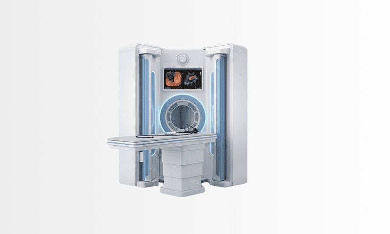
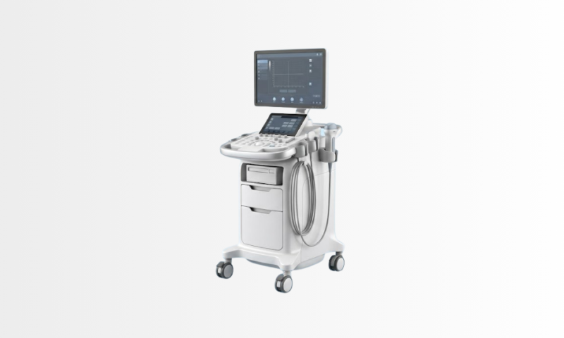
Available at these Hospitals

















What patients say about this unit
★★★★★From 2,400+ verified patient reviews“My gastroenterology workup, colonoscopy, imaging, labs, was compressed into three days without feeling rushed. Sedation was comfortable, results were discussed face to face, and the dietary plan was practical rather than generic.”
“After two inconclusive endoscopies at home, the gastroenterology team here combined capsule endoscopy with the right biopsies and found the answer. Treatment started the same week. Being believed was half the cure.”
Speak with our medical team
Share your case and our international patient team will guide you to the right specialist, free of charge.