

Neurosurgery
ACDF and cervical disc surgery, microdiscectomy, laminectomy and spinal fusion, deformity correction, plus brain tumour and skull base surgery, aneurysms, hydrocephalus, epilepsy and functional neurosurgery.
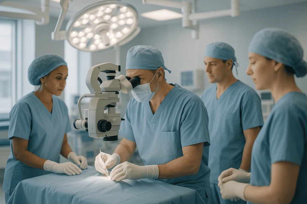
From a neck that has taken your grip to a scan you did not expect
Most people arrive with an arm or leg that has changed, a walking distance that keeps shrinking, or a brain scan taken for another reason. The work is deciding which of those needs an operation, and which does not.
Spine surgery
Discs, stenosis, instability and deformity, and the operations that answer each of them rather than all of them.
Brain and vascular surgery
Tumours, aneurysms and fluid disorders, planned by a board and operated with navigation and monitoring.
Functional neurosurgery
Stimulation and targeted procedures for movement disorders, epilepsy and facial pain that medicine no longer controls.
A hospital neurosurgical department, not a spine clinic
Spine surgery is easy to sell and easy to do for the wrong indication. The difference shows in who is turned away: a degenerate scan with symptoms that do not match it, back pain without instability, a herniation that is already resolving. A unit paid per operation is the wrong place to establish that.
It also shows when the finding is not the one anyone expected. Cord compression on a scan requested for neck pain, or a tumour behind a first seizure, needs neuroradiology, oncology and intensive care in the same building, not a referral letter to another city.
What we will not do
- Operate on a scan. Degeneration is treated when it explains the symptoms, not because it appears in a report.
- Add a fusion to a stable spine to make a decompression look more thorough.
- Promise that weakness or numbness will come back. Nerve recovery depends on how long the nerve was compressed, and that is said before surgery rather than after.
- Quote an operation before anyone has examined the patient it is quoted for.
- Treat non-surgical nerve disease as a surgical problem. Epilepsy on tablets, migraine and neuropathy belong to Neurology.
Neurosurgeons who lead this work
What actually happens, in order
Send the imaging, not the report
MRI and CT in DICOM format rather than printed pictures, plus standing flexion and extension X-rays for spinal cases, instability is a dynamic finding a lying-down scan cannot show.
Consultant review
Whether the scan explains the symptoms, whether the problem is a nerve root or the cord, and whether an operation is the right answer at all.
Board discussion where it applies
Every new brain tumour goes to a multidisciplinary board with neuroradiology, pathology and oncology before a treatment sequence is fixed.
Surgery day
Navigation, intraoperative neurophysiological monitoring and, where the anatomy calls for it, endoscopic or robotic assistance. Level confirmation before anything is removed.
Recovery and rehabilitation
Mobilisation from day one, a written rehabilitation plan, and the flying restriction explained in full before travel is booked.
Six things worth knowing first
Degeneration on a scan is not a diagnosis
Bulges, disc drying and facet wear are found in most people over fifty without symptoms. The question is which finding explains this patient, not what is abnormal.
Fusion is not the answer to back pain alone
Without instability or deformity, fusion has the least predictable outcome of any operation in this field. Decompression and fusion answer different problems.
Most sciatica settles without surgery
Disc fragments shrink over weeks to months in a large proportion of people. Surgery brings faster relief; by one to two years the gap narrows considerably.
Cord surgery stops decline first
In myelopathy the realistic goal is to halt deterioration. Some function returns, particularly when operated early, but it is not a restoration of what is lost.
Not every tumour needs removing now
Small meningiomas and acoustic neuromas are frequently watched with interval imaging, and many never grow enough to matter.
"Minimally invasive" is not a result
Smaller corridors mean an easier recovery, not a better decompression. An incomplete release through a small incision is the worse outcome.
Jump to what you came for
Quick answer
Neurosurgery is the medical specialty that diagnoses and treats disorders of the brain, spine, spinal cord, and peripheral nerves using both surgical and non-surgical approaches. At Acibadem in Turkey, neurosurgery covers evaluation, advanced imaging, treatment planning, and when needed procedures ranging from minimally invasive spine surgery to complex brain and nerve operations, followed by coordinated rehabilitation and follow-up.
What our neurosurgery unit covers, and who it is for
Neurosurgery is the surgical care of the brain, the spinal cord, the spine that houses it and the nerves that run from it. In practice that means two quite different populations meet the same department. One group arrives with a scan showing something inside the skull: a tumour, an aneurysm, fluid where it should not be. The other group arrives with a neck or back that has failed them: an arm that has gone weak, a leg that gives way, pain that has stopped responding to everything conservative. The operations are different, the risks are different, and the questions people ask are different. This page is written for both.
At Acıbadem International the work is organised into five strands, and knowing which one your problem belongs to is the fastest route to a useful answer.
- Spine surgerycervical, thoracic and lumbar degenerative disease, disc herniation, spinal stenosis, spondylolisthesis, deformity and fractures, from single-level decompression to instrumented fusion.
- Brain tumour and skull base surgerygliomas, meningiomas, metastases, pituitary and acoustic tumours, planned with the tumour board rather than by a single surgeon.
- Cerebrovascular neurosurgeryaneurysms and vascular malformations, treated either through the skull or from inside the vessel by the interventional route.
- Functional and epilepsy neurosurgerydeep brain stimulation, procedures for trigeminal neuralgia, and surgery for epilepsy that medicine has not controlled.
- Hydrocephalus and paediatric neurosurgeryshunts and endoscopic procedures for cerebrospinal fluid disorders, congenital malformations, and children’s spinal problems shared with pediatrics.
Two neighbouring specialties overlap with this one and it is worth being explicit about the border. Non-surgical brain and nerve conditions, stroke medicine, multiple sclerosis, epilepsy that is controlled on tablets, headache, neuropathy, belong to neurology. Spinal deformity in children and adults, joint and limb problems, and much sports-related back pain are shared with orthopedics; several spinal operations are performed by both specialties, and what matters is the surgeon’s experience with your specific operation rather than the label on the department door.
Who is this unit for? People holding a scan report they cannot decode. People who have been told they need spine surgery and want the reasoning examined before agreeing. People with a brain tumour diagnosis who want a plan built by a full team. And people whose previous spine operation did not solve the problem. If your question is “does this actually need surgery?”, the sections that follow were written for you.
Neurosurgeon vs neurologist: who does what
The neurosurgeon vs neurologist question comes up in almost every first consultation, and the distinction is simpler than it sounds. A neurologist diagnoses and treats diseases of the nervous system with medicines and other non-operative care: epilepsy, migraine, multiple sclerosis, Parkinson’s disease, neuropathy, most strokes. A neurosurgeon operates. Many conditions pass through both hands: an epilepsy that stops responding to drugs, a brain tumour that needs both surgery and oncological treatment, a Parkinson’s disease that reaches the stage where deep brain stimulation is considered.
Three practical consequences follow. First, a referral to a neurosurgeon is not a decision that you will be operated on; a large part of any neurosurgical clinic is telling people that surgery is not indicated. Second, the neurologist usually stays involved after an operation: the tablets, the seizure control, the rehabilitation plan do not stop because a surgeon was involved. Third, for spinal problems there is a third specialty in the picture: orthopedic spine surgeons train through a different route and perform many of the same operations, so a spine problem may reasonably be reviewed by either.
What a first neurosurgical consultation actually involves. The history comes first and carries more weight than most people expect: when the symptoms began, how they have changed, which movements or positions provoke them, what has already been tried. Then a neurological examination, power, sensation, reflexes, gait, coordination, and for spinal complaints the specific patterns that separate a nerve root problem from cord compression. Only then are the images reviewed, and they are reviewed against the examination. A scan that does not explain the symptoms is a common and important finding: it usually means the answer lies elsewhere, and operating on the wrong level or the wrong problem is how people end up in the failed-back category.
ACDF surgery (anterior cervical discectomy and fusion): the most common cervical operation
ACDF surgeryanterior cervical discectomy and fusion, removes a damaged cervical disc through a small incision at the front of the neck and replaces it with a spacer that allows the two vertebrae to fuse into one solid unit. It is the workhorse operation of cervical spine surgery, and it exists because of an anatomical fact: reaching the front of the spinal canal from behind would mean going through the spinal cord, while from the front the surgeon can reach the disc between structures that move safely aside.
The reason for the operation is almost never the disc itself. It is what the disc is pressing on. When a herniated or collapsed cervical disc narrows the space around a nerve root, the result is arm pain, numbness or weakness in a pattern that follows that nerve. When it narrows the canal around the cord, the result is cervical myelopathy, which behaves quite differently and is judged by a different set of rules.
Who actually needs ACDF surgery?
Surgery is considered when the imaging explains the symptoms and one of three situations applies: arm pain that has not settled after a reasonable course of non-operative treatment, weakness that is objectively present on examination, or spinal cord compression with signs of myelopathy. Neck pain alone, with a scan showing degeneration but no nerve compression, is generally not an indication, degenerative changes are so common in the general population by middle age that finding them proves very little on its own. Non-operative treatment first means physiotherapy, activity modification, medication prescribed by your doctor, and sometimes a targeted injection performed under imaging by pain management.
How the operation is performed, step by step
The patient is asleep under general anaesthesia. A transverse incision follows a natural skin crease at the front of the neck, usually two to three centimetres long. The surgeon works through the natural plane between the windpipe and oesophagus on one side and the carotid sheath on the other, no muscle is cut. Under the operating microscope the damaged disc is removed completely, along with any bone spurs narrowing the exit of the nerve root, until the dura and nerve are decompressed under direct vision. A spacer, a cage of titanium, PEEK polymer or bone graft, is placed in the empty disc space to restore its height, and a small plate with screws usually holds the segment still while fusion takes place. Fusion is what the body does over the following months; the metalwork only holds position while it happens.
How long does ACDF surgery take?
A single-level ACDF typically takes one to two hours of operating time, with additional time either side for anaesthesia and positioning; two- and three-level operations take proportionally longer. Most people spend one night in hospital, and some single-level cases are discharged the same day where the anaesthetic recovery has been straightforward. Those are typical ranges rather than promises: a re-operation, a heavily degenerated segment or an unexpected finding can change both figures, and your own surgeon’s estimate for your own anatomy is the one that counts.
ACDF recovery: what the weeks after surgery look like
ACDF recovery is usually easier than people fear at the incision and harder than they expect in the throat. Swallowing discomfort and a hoarse voice are common in the first days to weeks because the oesophagus and the nerve to the voice box were retracted; both settle in the large majority of cases. Arm pain caused by nerve compression often improves immediately, sometimes on waking. Numbness and weakness recover more slowly and less predictably, because a nerve that has been compressed for a long time takes months to recover and may not recover fully.
Walking starts the same day. A soft collar may be used for comfort for a short period, though many surgeons no longer use one after a single-level fusion with a plate. Desk work is commonly resumed within two to four weeks, physical work considerably later. Driving waits until you can turn your head freely and are off sedating medication. Lifting, pulling and overhead work are restricted for several weeks to months depending on the number of levels. The fusion itself continues to mature for six to twelve months, which is why smoking matters so much: nicotine measurably impairs bone healing, and it is the single most important thing a patient can change before this operation.
ACDF vs disc replacement: how the choice is made
The acdf vs disc replacement question is the most common one asked in cervical clinics, and it has a real answer rather than a preference. Fusion removes the disc and stops the segment moving. Cervical disc replacement removes the disc and installs an artificial joint that preserves motion. Motion preservation is attractive because a fused level transfers load to its neighbours, and adjacent-segment problems are a genuine long-term consideration.
What decides it is anatomy, not enthusiasm. Disc replacement requires a segment that still moves, facet joints in reasonable condition, alignment that is preserved, and absence of significant instability, infection, severe osteoporosis or advanced degeneration across multiple levels. Fusion remains the right operation where those conditions are not met, and where deformity needs correcting, where the segment is already unstable, or where a previous operation has changed the anatomy. A surgeon who offers only one of the two operations is describing their practice rather than your spine.
Does ACDF surgery work? An honest answer without numbers
You will not find a success-rate figure on this page. Published rates describe selected populations under particular definitions of success, and they travel badly to an individual with their own anatomy, duration of symptoms and general health. What can be said honestly is this: relief of arm pain from a decompressed nerve root is the outcome this operation achieves most reliably; neck pain responds less predictably, which is exactly why neck pain alone is a weak indication; recovery of weakness and numbness depends heavily on how long the nerve was compressed before surgery; and a proportion of people need further surgery years later, most often at an adjacent level. A surgeon who quotes you a single percentage without discussing which of those outcomes it refers to is not giving you information you can use.
Cervical disc replacement: motion preservation and its limits
Cervical disc replacementalso called cervical arthroplasty, removes the damaged disc exactly as a fusion does, then implants an artificial disc designed to keep the segment moving instead of letting it heal solid. The front-of-neck approach, the decompression and the recovery from the incision are near-identical to ACDF surgery; what differs is what is left behind in the disc space and what happens to the mechanics of the neck afterwards.
The argument for it is adjacent-segment disease. A fused level cannot move, so the levels above and below absorb more motion and more load, and over years some of them degenerate faster than they otherwise would. Preserving motion is intended to reduce that transfer. The argument is strongest in younger patients with single-level disease and an otherwise healthy neck, precisely the group in whom decades of adjacent-level loading lie ahead.
The limits are anatomical and non-negotiable. An artificial disc needs a mobile segment: if the level is already stiff, replacing the disc replaces nothing. It needs healthy facet joints, because the implant only addresses the front column and cannot help arthritic joints at the back. It needs reasonable bone quality, preserved alignment and no instability, infection or tumour. Severe multi-level degeneration, significant deformity and previous fusion at the same level generally take the patient back to fusion. Revision of a failed arthroplasty is a bigger undertaking than revision of a fusion, which is a fair thing to weigh at the outset rather than discover later.
What recovery adds. Because no bone needs to knit, the postoperative restrictions are usually lighter and movement is encouraged early. Anti-inflammatory medication, avoided after fusion because it can impair bone healing, is often permitted: a decision that belongs to the operating surgeon, not to a web page. Heterotopic ossification, in which bone forms around the implant and quietly stiffens the segment anyway, is a recognised long-term finding and one reason follow-up imaging continues for years.
Cervical myelopathy: when the cord, not the nerve, is compressed
Cervical myelopathy is compression of the spinal cord itself in the neck, usually from a combination of disc bulging, bone spurs, thickened ligament and, in some people, a canal that was narrow to begin with. It behaves nothing like a pinched nerve root and it is the single most important pattern for a spine clinic to recognise correctly, because the natural history is different: nerve root pain often settles by itself, whereas cord compression tends to progress in a stepwise way and recovered function is limited by how long the cord was compressed.
The presentation is deceptively quiet. Instead of severe pain, people describe clumsy hands, dropping cups, difficulty with buttons, handwriting that has deteriorated. Balance changes: a widened, careful gait, a hand on the wall in the dark. Numbness or a band-like feeling around the trunk. Neck pain may be mild or entirely absent, which is why the diagnosis is so often delayed. On examination the reflexes are brisk rather than reduced, and specific signs, Hoffmann’s, Babinski’s, a positive Romberg test, separate cord compression from a root problem within a minute of clinical testing.
How it is assessed. MRI shows the level and severity of compression and any signal change within the cord, which is a marker of established injury. Where MRI is not possible, CT myelography is the alternative. Nerve conduction studies do not diagnose myelopathy but help when the picture is mixed with a peripheral nerve problem. Because compression at more than one level is common, the whole cervical spine is imaged rather than the level that hurts.
What treatment involves. Established myelopathy with functional decline is one of the clearer surgical indications in spine practice; observation with regular review is reasonable only for mild, non-progressive disease. The operation is chosen by where the compression sits and how many levels are involved: an anterior approach, ACDF or corpectomy, for compression coming from the front at one or two levels; a posterior approachlaminectomy with fusion, or laminoplasty, which reconstructs the canal while preserving the bony arch, for multi-level compression, especially where alignment allows it. The realistic goal is stated plainly in every honest consultation: surgery is performed principally to stop deterioration. Some function returns, particularly in people operated on early, but the operation is not a restoration of what has already been lost.
Microdiscectomy and sciatica surgery: the lumbar disc operation
Microdiscectomy is the removal of the fragment of a herniated lumbar disc that is pressing on a nerve root, performed through a small incision with an operating microscope. It is the most common spinal operation in the world and the standard answer to disc-related sciatica surgery when non-operative treatment has failed. The word “micro” refers to the magnification and the size of the exposure, not to a lesser operation: the decompression achieved is the same as through a larger incision, with less muscle disruption on the way in.
The crucial framing is that most sciatica does not need an operation. A herniated disc fragment shrinks over weeks to months in a large proportion of people, and pain settles with it. That is why the standard advice is a period of non-operative care first, analgesia prescribed by your doctor, physiotherapy, maintained activity rather than bed rest, and in selected cases a nerve root injection performed under imaging. Surgery earns its place when that period fails, and the honest trade-off is well understood: operating brings faster relief of leg pain, while at one to two years the difference between operated and non-operated patients narrows considerably. What surgery does not do is change the natural history of the disc itself.
Who is a candidate, and who is not
The candidate has leg pain that dominates back pain, an MRI showing a herniation that matches the affected nerve root, and either a failed course of conservative treatment or progressive weakness. Poor candidates are people whose main complaint is back pain with a disc bulge on imaging, people whose scan does not match their symptoms, and people whose pain has been present for years without a clear neurological deficit. Two situations change the timetable rather than the indication: significant, progressing motor weakness, a foot that drops, a leg that gives way, and cauda equina syndrome, which is a surgical emergency and is treated as one wherever it presents.
Recovery, restrictions and the recurrence question
Leg pain frequently improves within hours of waking. Back soreness at the incision persists for a couple of weeks. Most people walk the same day, go home within twenty-four hours and return to desk work within two to four weeks; heavy manual work takes longer, and driving resumes when you can perform an emergency stop without hesitation. Bending, twisting and lifting are limited for the first few weeks while the annular defect in the disc heals over.
Recurrent herniation at the same level is the main long-term issue, and it happens because removing the fragment does not repair the hole it came through. Large annular defects carry a higher risk; so do smoking, obesity and early return to heavy lifting. A recurrence is not evidence that the first operation failed: it is the known behaviour of the injured disc, and it can be treated by repeat microdiscectomy or, where instability has developed, by fusion.
Herniated disc symptoms: reading the pattern
Herniated disc symptoms follow the nerve, not the spine. A lumbar disc pressing on the L5 root produces pain down the outer leg to the top of the foot with weakness of ankle and toe lift; an S1 root produces pain down the back of the leg to the sole with a reduced ankle reflex and weakness pushing off. In the neck, a C6 root gives pain to the thumb side of the forearm with a weak biceps, C7 to the middle finger with a weak triceps. This is why a competent examination can predict the level on the scan before the scan is opened, and why a mismatch between the two is such an important finding.
The vocabulary of radiology reports causes needless alarm. A bulge is a broad, symmetrical extension of the disc beyond its normal margin and is extremely common with age. A protrusion is a focal outpouching with the base wider than the dome. An extrusion has a dome wider than its base, and a sequestrated fragment has separated from the parent disc entirely. Counter-intuitively, the larger and more dramatic extrusions and sequestrations are the ones most likely to shrink substantially on their own, because the immune system resorbs displaced nuclear material. None of these words, on their own, is an indication for surgery.
Degenerative change reported as “disc desiccation”, “loss of disc height”, “annular fissure” or “Modic changes” describes the ageing spine. In population studies, a majority of people over fifty have several of these findings with no symptoms at all. Treating the report rather than the person is the commonest error in spinal care, and it is the reason a good clinic spends more time on examination than on the images.
Laminectomy, foraminotomy and lumbar decompression surgery
Laminectomy removes the lamina, the bony arch at the back of a vertebra, to enlarge a narrowed spinal canal and take pressure off the nerves inside it. It is the classic decompression operation, and in modern practice it has been joined by less destructive versions of the same idea. Foraminotomy enlarges the exit tunnel of a single nerve root, leaving the rest of the arch intact. Lumbar decompression surgery is the umbrella term, and the modern default is to remove exactly as much bone as the nerves need and no more, because every piece of bone and ligament removed is a piece of stability lost.
The distinction that matters clinically is between decompression alone and decompression with fusion. Removing bone relieves pressure; it does not stabilise anything. Where the segment is already slippingspondylolisthesisor where the decompression required is so extensive that it would destabilise the joint, fusion is added. Where the spine is stable and the problem is purely narrowing, adding fusion adds risk, cost and stiffness for no benefit. That judgement, made carefully, is one of the real differences between spine surgeons.
Unilateral approaches and tubular decompression
A canal can be decompressed from one side. Through a tubular retractor placed on one side of the midline, the surgeon works under the arch across to the opposite side, freeing both nerve roots while leaving the spinous process, the interspinous ligament and the muscle attachments of the far side untouched. The technique is often described as unilateral laminotomy for bilateral decompression, and it is the reason many patients now walk within hours of a decompression that once required a hospital week. It is not suitable for every anatomy, severe deformity, previous surgery with scarring, and instability all push the decision back toward an open approach.
Laminectomy recovery time: what is realistic
Laminectomy recovery time depends far more on the number of levels and the patient’s baseline than on the operation’s name. A single-level decompression in a fit person: walking the same day, home within one to two days, desk work in two to four weeks, walking distance improving over weeks as the nerves recover. A multi-level decompression in an older patient with other medical conditions: several days in hospital, a rehabilitation plan, and improvement measured over months. The dominant symptom of spinal stenosis, leg pain and heaviness on walking, usually improves first and most reliably. Numbness lags. Back pain may not change at all, because decompression was never aimed at it.
Recovery is an active process rather than a passive one. Walking is prescribed from day one and increased daily. Structured physiotherapy through physical medicine and rehabilitation restores the trunk strength lost during the months of limping before surgery, and it is the part of the plan patients most often skip and most often regret skipping.
Spinal stenosis symptoms and spinal stenosis surgery
Lumbar spinal stenosis is narrowing of the canal or the nerve exits, usually from a combination of thickened ligamentum flavum, enlarged facet joints and bulging discs: the accumulated result of decades of load. Spinal stenosis symptoms have a signature that separates them from almost everything else: neurogenic claudication. Pain, heaviness or numbness builds in the buttocks and legs after walking a certain distance, eases within minutes of sitting or bending forward, and returns at roughly the same distance next time. People describe leaning on a shopping trolley, walking uphill more easily than downhill, and cycling comfortably while being unable to walk to the corner, all because flexion opens the canal and extension closes it.
That pattern is worth knowing precisely, because vascular claudication from peripheral arterial disease produces similar leg pain but does not improve with bending forward and does not spare cycling; it is relieved simply by standing still. Distinguishing the two decides whether the answer lies in a spine clinic or a vascular one, and where both coexist, which is common in older patients, the assessment has to establish which is limiting the walking.
Spinal stenosis surgery is elective in the great majority of cases. Stenosis rarely damages nerves suddenly; the disability accumulates as walking distance shrinks. That makes the decision a quality-of-life judgement rather than an emergency: non-operative care, physiotherapy focused on flexion-based exercise, weight management, medication and sometimes epidural injections, is tried first, and decompression is offered when walking distance has fallen to a level the person is no longer willing to accept. What decompression reliably improves is walking distance and leg symptoms. What it does not reliably improve is chronic back pain, and a clinic that promises otherwise is overselling.
Spinal fusion and spondylolisthesis surgery
Spinal fusion joins two or more vertebrae into a single unit so that the segment between them stops moving. Metalwork, screws, rods, cages, holds the position; the fusion itself is biological, formed over months as bone grows across the segment. The list of legitimate reasons is short and worth stating, because fusion is the operation most often performed for the wrong reason: instability, deformity that needs correcting, fracture, tumour, infection, and decompression so extensive that stability would otherwise be lost.
Spondylolisthesis: the slip that decides the operation
Spondylolisthesis surgery is the clearest of those indications. Spondylolisthesis is one vertebra slipping forward on the one below, either through a defect in the bony bridge (isthmic, often dating from adolescence) or through worn facet joints (degenerative, typically after fifty). Where the slip is stable and the symptoms are tolerable, no operation is needed. Where it is progressing, where the nerves are compressed, or where the slip moves on flexion and extension X-rays, decompression alone risks making the segment more unstable, so decompression and fusion are performed together.
Approaches, in plain terms. PLIF and TLIF reach the disc space from behind, through or around the nerve structures. ALIF approaches from the front through the abdomen, giving a large graft surface and good correction of alignment but requiring an approach past the great vessels, usually with a vascular or general surgeon present. LLIF and OLIF come in from the side, avoiding both the back muscles and the abdominal contents, with their own nerve-related considerations at particular levels. No approach is universally superior; each is chosen for the level, the anatomy, the alignment goal and the surgeon’s experience.
The honest counterweight. Fusion trades motion for stability. That trade is worth it when instability is the problem and a poor bargain when it is not. Non-union, where the bone does not knit, is a recognised complication, strongly associated with smoking, and it is the main reason surgeons are so insistent about nicotine before elective fusion. Adjacent-segment degeneration develops in a proportion of patients over years. And fusion performed for back pain alone, with no instability and no deformity, has the least predictable outcome of any operation in this chapter.
Minimally invasive spine surgery and endoscopic spine surgery
Minimally invasive spine surgery describes a family of techniques that reach the same targets through smaller corridors: tubular retractors that split muscle instead of stripping it from bone, percutaneous screws placed through stab incisions under imaging, and interbody cages inserted through the side of the body. The advantages are real and measurable, less blood loss, less postoperative pain, shorter hospital stays, faster mobilisation. The advantage that is sometimes implied but not delivered is a better long-term result: at one to two years, a well-performed open operation and a well-performed minimally invasive one aim at the same destination. The gain is in the journey.
Endoscopic spine surgery takes the idea furthest. Working through a cannula a few millimetres wide with a rod-lens endoscope and continuous irrigation, the surgeon removes a disc fragment or decompresses a foramen through an incision that needs a single stitch. Selected cases are done under local anaesthesia with sedation, which matters for patients whose general health makes general anaesthesia risky. The limits are equally clear: the working corridor is small, the anatomy must suit it, revision cases with scar tissue are difficult, and the learning curve is long enough that experience with the specific technique is a fair question to ask a surgeon.
The honest summary for a patient comparing quotes: “minimally invasive” on a brochure means very little by itself. What matters is whether the specific operation proposed for your specific problem can be done adequately through that corridor, and whether the decompression achieved is complete. An incomplete decompression through a beautiful small incision is a worse outcome than a complete one through a larger scar.
Reading your spine MRI report: what the words actually mean
More people arrive at a spine clinic frightened by a report than by their symptoms. Radiology language is precise, unfamiliar and written for another doctor, and read cold it can make an ordinary middle-aged spine sound like a catastrophe. This section translates the terms that appear most often, in the order they usually appear in a lumbar or cervical MRI report.
Disc terminology, in plain language
Disc desiccation means the disc has lost water content and appears darker on the scan. It is the earliest and most universal sign of disc ageing; by the sixth decade it is present in the majority of people, most of whom have no back pain at all. Loss of disc height is the mechanical consequence of the same process. Annular fissuresometimes reported as a high-intensity zone, describes a tear in the tough outer ring; it may be a source of pain and may equally be an incidental finding, which is why it cannot be treated as a diagnosis on its own. Schmorl’s node is disc material pushed into the vertebral body above or below, usually asymptomatic. Modic changes describe signal alteration in the bone next to a degenerated disc, graded I to III; type I is the one most often associated with pain, though the association is far from absolute.
Canal, foramen and facet terminology
The report will grade narrowing in three separate places, and they mean different things clinically. Central canal stenosis narrows the space around the whole nerve bundle and produces the walking-limited pattern of neurogenic claudication. Lateral recess stenosis narrows the space where a single root turns to leave, and produces pain in that root’s territory. Foraminal stenosis narrows the exit tunnel itself, often worsened by standing and extension, and is the pattern most likely to need a foraminotomy rather than a central decompression. Facet arthropathy describes wear in the small paired joints at the back of each segment; ligamentum flavum hypertrophy describes the thickening of the ligament that forms the back wall of the canal, and it contributes substantially to stenosis in older spines. Synovial cyst is a fluid-filled outpouching from a worn facet joint that can compress a nerve root on its own.
Alignment, instability and the words that change the operation
Spondylolisthesis is a forward slip and is graded I to IV by how far the vertebra has moved. Retrolisthesis is a backward slip. Spondylolysis is a defect in the bony bridge that allows a slip to occur, classically in adolescent athletes. Scoliosis is a lateral curve, kyphosis a forward one, lordosis the normal inward curve of the neck and lower back, and the loss of lumbar lordosis, often described as “flattening”, is a finding with real consequences for standing balance. Crucially, an MRI is taken lying down, when the spine is unloaded. Instability is a dynamic phenomenon, and the only way to see it is with standing flexion and extension X-rays. A report that says nothing about instability has not excluded it.
Cord signal, syrinx and the findings that change urgency
In the cervical and thoracic spine the report will comment on the cord itself. Cord signal changeincreased signal on T2 sequences, indicates injury to the cord from compression and is one of the findings that shifts a case from watchful waiting toward surgery, because it marks damage that has already occurred. Syrinx describes a fluid cavity within the cord, seen in Chiari malformation, after trauma and with some tumours. Cord compression with effacement of the cerebrospinal fluid space means the protective fluid layer around the cord has been squeezed out. And marrow replacement or abnormal enhancement in a vertebral body raises the question of tumour or infection rather than degeneration, and moves the assessment into a different pathway entirely.
Why the report is never the whole answer
The single most useful thing to understand about spinal imaging is how common abnormal findings are in people without symptoms. Disc degeneration, bulges, facet arthritis and even frank herniations are found routinely in volunteers who have never had back pain, and their prevalence rises steadily with each decade. This is not a reason to distrust imaging; it is the reason imaging is interpreted alongside the examination rather than instead of it. The question a surgeon asks is never “what is abnormal on this scan?” but “which abnormality on this scan explains this person’s symptoms?”, and when nothing does, that answer is itself important, because it redirects care away from an operation that would not have worked.
MRI, CT, myelogram and angiography: which test answers which question
MRI is the workhorse of neurosurgery because it shows soft tissue: discs, nerves, spinal cord, brain, tumours and inflammation. It uses no ionising radiation. Its limitations are practical: it is slow, it is noisy, it is difficult for people with severe claustrophobia, and it is contraindicated or requires special protocols with certain implanted devices, which is why the safety questionnaire before a scan matters. Contrast (gadolinium) is added when tumour, infection or scar tissue is in question, because these enhance while normal tissue does not.
CT shows bone with a clarity MRI cannot match, and it is fast, which is why it is the first test after head injury and in an unstable patient. In spinal work it defines fracture anatomy, bone quality, the position of implants and the exact shape of bony spurs. CT myelography, in which contrast is injected into the fluid around the cord before scanning, is the alternative when MRI is impossible or when metal from previous surgery obscures the anatomy; it outlines the nerve structures indirectly and remains genuinely useful in revision cases.
Catheter angiographythe injection of contrast directly into the cerebral vessels, remains the reference standard for aneurysms and vascular malformations, showing flow dynamics that non-invasive tests can miss, and it is the same platform on which coiling is performed. CT angiography and MR angiography are non-invasive alternatives used for screening and follow-up. PET answers metabolic questions in tumour work, distinguishing recurrence from treatment effect, or locating a primary tumour when a brain metastasis is the first sign of cancer. Nerve conduction studies and EMG are not imaging at all; they test how nerves and muscles actually function, and they answer questions imaging cannot, such as whether an abnormal-looking nerve is genuinely the source of weakness.
What comes before surgery: non-operative spine care
The majority of people who see a spine surgeon do not have an operation, and the treatments that fill that gap deserve more space than they usually receive. They are not a waiting room for surgery; for most degenerative conditions they are the treatment.
Structured physiotherapy is the backbone. What it targets depends on the diagnosis, extension-based programmes for some disc problems, flexion-based work for stenosis, core and hip strengthening for instability-related pain, nerve gliding for radicular symptoms. What separates effective from ineffective is specificity and progression, not the number of sessions. Activity modification means changing how you load the spine, not stopping moving; prolonged bed rest worsens outcomes and is no longer recommended for acute back pain.
Medication belongs to the prescribing doctor. Simple analgesia, anti-inflammatories, short courses of muscle relaxants and agents aimed specifically at nerve pain each have a place, and each has limits and side effects that make the choice individual. Image-guided injections, performed by pain management or radiology, serve two purposes that are worth separating: therapeutic, to reduce inflammation around an irritated nerve root and buy time for natural resolution; and diagnostic, to test whether a specific structure is generating the pain by numbing it temporarily. A diagnostic block that abolishes the pain for the expected duration is a genuinely useful piece of information before considering surgery on that level.
Radiofrequency ablation of the small nerves supplying arthritic facet joints can give months of relief in carefully selected patients identified by diagnostic blocks. Weight management, smoking cessation and treatment of osteoporosis alter the trajectory of degenerative spinal disease more than any single intervention on this list, and they remain relevant whether or not an operation eventually happens, because they determine how well anything that is done will hold.
Robotic spine surgery and spinal navigation
Robotic spine surgery is not a robot performing an operation. It is a computer-controlled arm that holds a guide in a precisely planned trajectory so the surgeon can place a screw exactly where the plan says it should go. The plan is built beforehand from a CT scan: every screw’s entry point, angle, diameter and length is chosen on a three-dimensional model, then registered to the patient on the operating table. The surgeon does the surgery; the arm removes hand tremor and guesswork from one specific step of it.
Navigation is the same idea without the arm. A reference frame is fixed to the spine, an intraoperative scan registers the anatomy, and instruments tracked by an optical camera appear on screen in real time relative to the patient’s own images. In practice a modern deformity or instrumentation case uses navigation as standard, with robotics added where the geometry is demanding: severe deformity, revision anatomy, obesity that limits fluoroscopy, or percutaneous placement where landmarks are not visible.
What it changes and what it does not. Accuracy of screw placement improves and radiation exposure to the surgical team falls, because fewer live fluoroscopy shots are needed. What technology cannot supply is the decision, whether to operate, at which levels, with which alignment goal. A perfectly placed screw in a poorly chosen construct is still a poorly chosen construct. This is the honest framing every patient should hear when a hospital advertises equipment: the machine improves execution, and execution was rarely the reason a spine operation failed.
Scoliosis surgery: curves in children and adults
Scoliosis surgery answers two quite different problems that share a name. In adolescents, the issue is a growing curve: idiopathic scoliosis is monitored by curve magnitude and remaining growth, braced within a defined range while the skeleton is still immature, and considered for surgery when the curve passes roughly forty to fifty degrees or continues to progress despite bracing. The operation corrects and fuses the curve, and the goal is a balanced, stable spine into adult life. In adults, the issue is usually not the curve’s cosmetic appearance but what the curve is doing to the nerves and to standing balance: leg symptoms from stenosis within the deformity, and the exhausting forward or sideways lean of sagittal imbalance.
Adult deformity surgery is among the largest operations in this field, and honesty about its scale is part of informed consent: long constructs, significant blood loss, several hours of operating, intensive care in some cases, and a complication rate meaningfully higher than that of a single-level decompression. It is offered when the disability is severe and the alternatives have been exhausted, and it is planned with alignment parameters measured on full-length standing radiographs rather than by eye. For children and adolescents the pathway is shared with orthopedics and paediatric spine specialists, who between them see the volume that makes this work safe.
Two practical points patients ask about constantly. Growth: fusing a growing spine is avoided where possible, and growth-friendly techniques exist for very young children with severe curves. Motion: a fused thoracic curve costs surprisingly little functional movement, because most trunk rotation comes from segments that are usually left free, but a fusion extending into the lumbar spine is felt, and that is discussed before rather than after.
Kyphoplasty and vertebral compression fractures
Kyphoplasty treats a collapsed vertebra, most often caused by osteoporosis and sometimes by tumour. Through a needle placed into the vertebral body, a balloon is inflated to create a cavity and partially restore height, then bone cement is injected to stabilise it. Vertebroplasty is the same idea without the balloon: cement injected directly into the fractured bone. Both are performed under imaging guidance, usually with sedation or light anaesthesia, and most patients go home the same or next day.
The nuance that gets lost in marketing is patient selection. Most osteoporotic compression fractures heal with time, analgesia and mobilisation; the trials that showed the least benefit for cement were those that included patients whose pain was already settling. The strongest case for intervention is a recent fracture with severe, focal, movement-related pain that has not responded to a reasonable course of conservative management, with oedema on MRI confirming the fracture is still active. Old, healed fractures do not respond to cement, and identifying which fracture on a scan is the painful one is precisely what the MRI is for.
Beyond the fracture lies the more important issue: the bone. A fragility fracture is a diagnosis of osteoporosis until proven otherwise, and a spine that has broken once will break again if nothing changes. Bone density assessment and medical treatment of osteoporosis, coordinated with endocrinology, do more for the next ten years than the cement does for the next ten weeks. Treating the fracture without treating the bone is the commonest failure of care in this group.
Failed back surgery syndrome and spinal cord stimulation
Failed back surgery syndrome is an unhelpful name for a real problem: persistent pain after technically completed spinal surgery. The name implies the surgeon failed, when the commonest explanations are different: the operation addressed a finding that was not the source of pain, the nerve had been compressed too long to recover, epidural scarring formed around the root, a segment adjacent to the fusion has now degenerated, or the original problem has recurred. Untangling which of these applies is the entire task, and it starts with a fresh history and examination rather than with a repeat operation.
Investigation typically means MRI with contrast, contrast distinguishes scar tissue, which enhances, from recurrent disc, which does not, plus flexion-extension films for instability and, where infection is possible, inflammatory markers. Diagnostic injections have a role in identifying whether a specific root or facet joint is generating the pain. Re-operation is reasonable when a clear, correctable structural cause is found. Re-operating on scar tissue alone is not: scar reliably reforms.
Spinal cord stimulator: trialled before it is implanted
Where no correctable cause exists, the direction of care changes from surgical to functional, coordinated with pain management. A spinal cord stimulator delivers mild electrical pulses through leads in the epidural space to modify pain signalling. Its defining feature is that it is trialled before it is implanted: leads are placed temporarily for several days, and only patients who obtain meaningful relief during the trial proceed to a permanent system. That trial is the honest part of the technology, and it belongs to a multidisciplinary programme that also addresses conditioning, sleep, mood and medication, because a device alone rarely rebuilds a life narrowed by years of pain.
Cauda equina syndrome: the spinal emergency
Cauda equina syndrome is compression of the bundle of nerve roots at the bottom of the spinal canal, most often by a large central disc herniation, and it is the one spinal condition where hours matter. The nerves involved supply the bladder, the bowel, the sexual organs and the saddle area, and the damage they sustain under sustained compression is frequently permanent. The pattern is distinctive: numbness in the saddle area, the inner thighs, buttocks and perineum, with difficulty starting or feeling urination, loss of the sensation of a full bladder, faecal incontinence or loss of anal sensation, sexual dysfunction of sudden onset, and often bilateral sciatica or leg weakness.
It is a surgical emergency, diagnosed by urgent MRI and treated by urgent decompression in hospital; the volume of the disc fragment removed is the same as in a routine microdiscectomy, and the difference between a good and a poor outcome is time rather than technique. Bladder function that has already been lost recovers unpredictably, which is why the whole system around this diagnosis is built for speed.
Two related conditions share the same logic. Compression of the spinal cord itself by tumour, infection or fracture, metastatic cord compression above all, behaves in the same time-critical way and is managed jointly with medical oncology and radiation oncology. Spinal epidural abscess, seen most often in people with diabetes, intravenous drug use or recent bacteraemia, combines fever, spinal pain and neurological deficit, and needs drainage and antibiotics rather than observation.
Head injury, chronic subdural haematoma and traumatic brain injury
Head injury spans an enormous range, from a knock that produces a brief headache to a catastrophic brain injury, and neurosurgery is involved in a narrow but critical band of it. The initial assessment everywhere follows the same logic: level of consciousness, pupils, focal deficits, and CT imaging when specific criteria are met. Most people with a mild head injury have a normal scan and recover fully; the purpose of imaging is to find the minority whose bleeding will progress.
Chronic subdural haematoma deserves particular attention because it is common, treatable and frequently missed. Weeks after an injury so minor that the patient may not recall it, blood slowly collects between the brain and its covering, and because it accumulates gradually the brain accommodates it. The presentation is therefore not dramatic: confusion, personality change, unsteadiness, headache, or a weakness that develops over days and is mistaken for a stroke or for dementia. It is more common in older people, in those taking anticoagulants, and after alcohol excess. Treatment is surgical drainage, usually through small burr holes under local or general anaesthesia, and the recovery in an otherwise well patient is often striking. Middle meningeal artery embolisation, performed by the interventional team, has emerged as an adjunct or alternative in selected cases.
Acute traumatic bleedingextradural haematoma from a torn artery, acute subdural haematoma from bridging veins, contusions within the brain, is a different, time-critical entity managed in a trauma centre with neurosurgical and intensive care support. Decompressive craniectomy, in which a large piece of skull is removed and left off to give the swelling brain room, is used for uncontrollable intracranial pressure; the bone is replaced in a later operation once swelling resolves. Recovery from significant traumatic brain injury is measured in months and years and belongs as much to rehabilitation and neurology as to surgery.
Surgery in stroke and intracerebral haemorrhage
Most strokes are treated medically and by interventional techniques rather than by open surgery, but neurosurgery holds several defined roles. In ischaemic stroke, a large infarction of the middle cerebral artery territory can swell dangerously in the days after the event; decompressive hemicraniectomy relieves that pressure and is a life-saving operation with a narrow time window and a carefully weighed discussion about the disability that may follow. Cerebellar infarction can compress the brainstem and block cerebrospinal fluid drainage, and is managed surgically for the same mechanical reasons.
In intracerebral haemorrhage, surgery is considered when a clot is superficial and large enough to be causing pressure, when a cerebellar haemorrhage threatens the brainstem, or when an underlying lesion, an aneurysm, a vascular malformation, a tumour, needs treating in its own right. Minimally invasive evacuation techniques through a small corridor are used in selected cases. Deep haemorrhages in the basal ganglia are frequently managed medically, because the surgical route to them costs more brain than the clot does.
Endovascular thrombectomy for large-vessel occlusion is performed by the neurointerventional team on the same platform used for aneurysm treatment, within a defined time window and with imaging criteria that identify who will benefit. It sits beside neurosurgery rather than within it, which is precisely why a centre that offers both under one roof shortens the chain of decisions in the hours that matter most.
Paediatric neurosurgery
Children are not small adults in this specialty, and the conditions differ as much as the anatomy. Hydrocephalus in infants presents with a rapidly enlarging head, a bulging fontanelle, downward-deviating eyes and feeding difficulty, because the skull can still expand before pressure symptoms appear. Treatment follows the same logic as in adults, shunt or endoscopic third ventriculostomy, with the added consideration that a shunt placed in infancy will be part of that person’s life for decades and will need revising.
Craniosynostosis is the premature fusion of one or more skull sutures, producing a characteristic head shape and, in multi-suture forms, the risk of raised pressure and impaired brain growth. Correction is performed with plastic and craniofacial surgeons, either through endoscopic techniques in young infants followed by helmet therapy, or through open remodelling later. Spinal dysraphismspina bifida and its milder tethered-cord variants, is managed with an eye on function rather than appearance: the goal is preserving bladder, bowel and leg function, which means surgery is timed by neurological monitoring rather than by the finding alone.
Paediatric brain tumours differ from adult ones both in type and in location, with a higher proportion arising in the posterior fossa and presenting with unsteadiness, morning vomiting and headache. They are treated within a paediatric oncology framework, and the long-term picture includes the effects of treatment on a developing brain, which is why radiation strategies in children are approached with particular caution and why long-term neurocognitive follow-up is part of the plan rather than an afterthought. Care is shared throughout with pediatrics.
Skull base surgery: the corridors under the brain
The skull base is the floor on which the brain rests: a complex plate of bone pierced by every cranial nerve and every major vessel entering or leaving the head. Tumours here are often benign but are difficult precisely because of what surrounds them: meningiomas of the petroclival region, chordomas along the midline, paragangliomas at the jugular foramen, and tumours extending upward from the sinuses. Access requires either a large, carefully planned open approach or an endoscopic corridor through the nose, and increasingly a combination of both in the same operation.
Two principles govern this work. The first is teamwork: skull base surgery is routinely performed jointly by neurosurgeons and ENT surgeons, with head and neck, ophthalmology, plastic and vascular colleagues involved depending on the anatomy: a genuine multi-specialty operation rather than a courtesy. The second is that function outranks completeness. A remnant of a benign tumour left deliberately on a cranial nerve, and then watched or treated with radiosurgery, is a better outcome than a complete removal that costs the patient their swallowing, their facial movement or their vision. Reconstruction of the skull base after resection, with local flaps of tissue, fat grafts and sometimes free tissue transfer, is what prevents cerebrospinal fluid leaking afterwards, and it is a substantial part of the operation rather than its closing act.
Less common lesions with well-defined answers
Several conditions appear rarely in any one clinic but have clear, established management, and patients researching them often find only fragments. Hemifacial spasminvoluntary twitching that begins around one eye and spreads down the same side of the face, is usually caused by a blood vessel compressing the facial nerve at the brainstem, and microvascular decompression addresses it in the same way it addresses trigeminal neuralgia. Cavernoma is a cluster of abnormal thin-walled vessels that may bleed in small amounts; many are watched, and surgery is considered for repeated haemorrhage, seizures that resist medication, or an accessible location after a significant bleed.
Colloid cyst sits at the roof of the third ventricle and can intermittently block cerebrospinal fluid flow, causing positional headaches, and is removed endoscopically or microsurgically when symptomatic or when the anatomy suggests risk. Arachnoid cyst is a fluid collection between the brain’s coverings, usually congenital and usually asymptomatic, most require nothing at all, and the main clinical skill is resisting the temptation to treat a finding rather than a patient. Pineal region lesions present with hydrocephalus and characteristic eye movement changes, and are approached with biopsy and fluid diversion before definitive treatment because tumour type varies so widely. Syringomyeliaa fluid cavity in the cord, is treated by addressing its cause, most often a Chiari malformation or post-traumatic scarring, rather than by draining the cavity itself.
Brain tumor surgery (brain tumour surgery): what the operation is for
Brain tumor surgery has three separate purposes, and knowing which one applies to a given patient explains most of what follows. The first is diagnosis: tissue is needed because treatment differs completely between tumour types, and modern classification depends on molecular markers that only tissue can provide. The second is relief of pressure: a mass in a closed box causes symptoms by displacing and compressing what is around it, and removing bulk relieves that directly. The third is disease control: for many tumour types the extent of resection is one of the few factors under the surgeon’s influence that correlates with outcome.
What is never promised is completeness where anatomy forbids it. Tumours that infiltrate rather than displace, diffuse gliomas above all, do not have an edge that can be cleanly followed, and tissue that looks normal at the margin already contains tumour cells. Tumours sitting in or against eloquent territory, speech, motor, vision, the brainstem, impose a limit that has nothing to do with the surgeon’s ambition: removing the last few millimetres can cost a function the patient values more than the millimetres are worth. A plan that states this openly before surgery is a better plan than one that discovers it afterwards.
Brain tumor symptoms and how they are investigated
Brain tumor symptoms arise in three ways. Raised pressure produces headache that is worse in the morning or on lying flat, nausea, vomiting and visual blurring. Local irritation produces seizures: a first seizure in an adult is investigated with imaging for exactly this reason. Local pressure produces focal deficits that map to the site: weakness on one side, speech disturbance, visual field loss, personality or memory change in frontal and temporal locations, unsteadiness in the cerebellum. Slow-growing tumours can reach a considerable size before any of this appears, because the brain accommodates gradual change remarkably well.
Investigation is MRI with contrast, supplemented as needed by advanced sequences: perfusion imaging to assess vascularity, spectroscopy for tissue chemistry, diffusion tensor imaging to map white matter tracts near the planned corridor, and functional MRI to locate language and motor areas relative to the tumour. Where a metastasis is suspected, imaging of the body follows to find the primary. Where lymphoma is a possibility, steroids are ideally withheld until tissue is obtained, because they can shrink the lesion enough to make the biopsy uninformative.
Why the tumour board decides, not the surgeon
Every new brain tumour at Acıbadem is discussed by a multidisciplinary board, neurosurgery, neuroradiology, neuropathology, radiation oncology and medical oncologybefore a treatment sequence is fixed. The reason is structural rather than ceremonial: the best first step for a given tumour is often not surgery, and the person best placed to judge that is not always a surgeon. A small metastasis in an eloquent area may be better treated with radiosurgery; a suspected lymphoma needs a biopsy, not a resection; a low-grade glioma in a young patient may be watched for a period under close imaging. A single opinion cannot cover that ground.
Craniotomy: opening and closing the skull
Craniotomy is the temporary removal of a piece of skull to reach the brain, replaced and fixed at the end of the operation with small plates and screws. The location and size of the opening are chosen for the shortest safe corridor to the target rather than for the size of the lesion. Modern practice has made the openings considerably smaller: navigation shows the surgeon exactly where the lesion sits relative to the surface, so the bone flap is planned to the target rather than to a landmark.
Inside, the work is done under an operating microscope or exoscope with microsurgical instruments, ultrasonic aspirators to remove tumour bulk and bipolar diathermy for haemostasis. Intraoperative neurophysiological monitoring tracks the function of motor pathways and cranial nerves continuously, giving a warning before an injury becomes permanent. Intraoperative ultrasound or, where available, intraoperative MRI answers the surgeon’s most difficult question in real time: is there tumour left that can safely come out?
Craniotomy recovery begins in a monitored bed, often intensive care for the first night, with regular neurological observation. Headache at the wound, jaw stiffness if the temporalis muscle was involved, and profound fatigue for several weeks are all expected. Most people are walking within a day or two. Driving and flying are restricted for a defined period that depends on the operation and on whether seizures occurred; anti-seizure medication is prescribed and adjusted by the treating team. Fatigue is the symptom patients are least prepared for, and it commonly outlasts the wound by months.
Awake craniotomy: operating with the patient talking
Awake craniotomy is used when a lesion sits in or beside areas that carry language or fine motor function, where no scan can define the border precisely enough. The patient is asleep for the opening and closing and awake for the resection, with the scalp anaesthetised locally. During the resection a neuropsychologist runs continuous testing, naming objects, counting, moving a hand, while the surgeon stimulates points on the cortex and along the tracts. A point that reliably disrupts speech when stimulated marks a boundary that is not crossed.
It sounds far more alarming than patients report it to be, and it exists for one reason: it maps function in the individual brain rather than the average brain. Language areas vary between people, and shift when a slow-growing tumour has pushed them. The technique requires a patient able to cooperate and a team practised at it; it is not offered for every lesion, and it is not a marketing feature but a tool with a specific indication.
Meningioma surgery: the tumour that is often watched
Meningioma surgery is one of the clearest examples of the principle that finding a tumour and needing an operation are different things. Meningiomas arise from the coverings of the brain, are usually slow-growing and benign, and are found incidentally with increasing frequency as scanning becomes routine. A small, asymptomatic meningioma in an older patient is very often best watched with interval imaging: many never grow enough to matter, and observation carries none of the risks of surgery.
Surgery is considered when the tumour is symptomatic, when it is demonstrably growing on serial scans, when it causes oedema in the surrounding brain, or when its position means that later growth would be far harder to treat. Because meningiomas displace rather than infiltrate, complete removal is often achievable, and the completeness of removal, including the dural attachment, is the main determinant of recurrence. Location dictates difficulty: a convexity meningioma over the surface is a very different operation from one at the skull base wrapped around cranial nerves and arteries, where a planned partial removal followed by radiosurgery to the remnant is often the wiser strategy.
Pituitary tumor surgery: through the nose, not the skull
Pituitary tumor surgery is usually performed endoscopically through the nostril and the sphenoid sinus, with no incision on the head at all. The endoscope provides a wide, illuminated view of the sella; the tumour is removed from within, and the skull base is reconstructed to prevent cerebrospinal fluid leaking into the nose. It is done jointly by a neurosurgeon and an ENT surgeon in most high-volume centres, and it is one of the operations where a shared, practised team makes a measurable difference.
The indications sort into three groups. Tumours that press: a lesion large enough to compress the optic chiasm classically takes the outer halves of both visual fields, and visual loss is a strong indication to operate. Tumours that secrete: growth hormone excess causing acromegaly, ACTH excess causing Cushing’s disease, and rarer secreting tumours are treated surgically when medical therapy is not the first-line answer. And prolactinomas, which are the exception: they typically respond so well to medical treatment prescribed by endocrinology that surgery is reserved for the minority who cannot tolerate or do not respond to it.
What follows the operation. Hormone replacement is assessed carefully in the days afterwards, because the pituitary controls thyroid, adrenal and other axes; temporary or permanent diabetes insipidus, causing thirst and large volumes of dilute urine, is a recognised outcome and is managed medically. Nasal congestion and crusting last several weeks. Vision often begins to improve within days when it was recently lost, and less predictably when it was lost long ago. Long-term endocrine follow-up is not optional: this is a shared disease between two departments for years, not a single operation.
Acoustic neuroma and acoustic neuroma surgery
Acoustic neuromavestibular schwannoma in current terminology, is a benign tumour growing from the sheath of the balance nerve as it runs with the hearing nerve and the facial nerve through a narrow bony canal to the inner ear. Because three critical nerves share that corridor, a tumour of a few millimetres can announce itself early: one-sided hearing loss, one-sided tinnitus, unsteadiness. Sudden or asymmetric hearing loss in one ear is the classic reason an MRI of the internal auditory canals gets requested, and it is why this tumour is usually found by an ENT clinic rather than a neurosurgical one.
There are three legitimate management options and the choice is genuinely balanced. Observation with serial MRI suits small tumours, older patients and tumours that have shown no growth, many grow very slowly or not at all. Stereotactic radiosurgery controls growth in a high proportion of small and medium tumours in a single session, without an incision. Acoustic neuroma surgery removes the tumour and is favoured for larger lesions, those pressing on the brainstem, and those causing hydrocephalus. Each option is weighed against hearing that remains useful, facial nerve function, the patient’s age and the tumour’s growth history, and it is one of the few decisions where a second opinion is genuinely standard practice rather than a sign of distrust.
The surgical approach, translabyrinthine, retrosigmoid or middle fossa, is chosen for tumour size, position and whether hearing preservation is realistic. Facial nerve function is monitored continuously throughout the operation, and preserving it takes priority over removing the last fragment of tumour adherent to it; a planned small remnant, watched or treated later with radiosurgery, is a deliberate and defensible strategy. Recovery includes a period of imbalance while the brain compensates for the loss of one balance nerve, which vestibular rehabilitation accelerates considerably.
Gliomas and glioblastoma treatment: honesty about what surgery can do
Gliomas arise from the supporting cells of the brain and span a wide range of behaviour, from slow-growing low-grade tumours in young adults to glioblastoma treatment, which addresses the most aggressive of them. Classification now depends on molecular markers as much as on appearance under the microscope, IDH mutation status, 1p/19q co-deletion, MGMT promoter methylation, because those markers predict behaviour and response to treatment far better than shape alone. This is one reason tissue matters and why biopsy is not a formality.
The standard sequence for glioblastoma is maximal safe resection, followed by radiotherapy with concurrent and then adjuvant chemotherapy, coordinated between neurosurgery, radiation oncology and medical oncology. “Maximal safe” is the operative phrase and it is not a hedge: the extent of resection is associated with outcome, and so is the neurological function the patient keeps. Fluorescence-guided surgery, in which the patient takes a compound that makes tumour tissue glow under a specific wavelength of light, helps the surgeon see the edge that white light does not show.
What this page will not do is imply a trajectory that the evidence does not support. Glioblastoma is not curable with current treatment; the aim is length and quality of life, and both are worth pursuing seriously. Low-grade gliomas behave very differently, often over many years, and in selected cases are observed rather than treated immediately. Clinical trial participation is discussed openly where a relevant trial exists, because for this disease the standard of care and the frontier of research sit unusually close together.
Brain metastases: the commonest brain tumour
Secondary tumours that have spread from elsewhere, most often lung, breast, melanoma, kidney and colorectal primaries, outnumber primary brain tumours several times over. They tend to be well-circumscribed, sit at the junction between grey and white matter, and are frequently multiple. That makes their management a different discipline from primary tumour surgery: the question is rarely “can this be removed?” but “what does this patient’s whole disease need?”
Surgery is favoured for a single large lesion causing pressure, for a lesion in the posterior fossa threatening the fluid pathways, and when tissue is needed to establish the primary or its molecular profile. Radiosurgery treats several small lesions in one or a few sessions and has largely displaced whole-brain radiotherapy as the first choice where the number of lesions allows it. Systemic therapy has changed the picture substantially: several modern targeted agents and immunotherapies cross into the brain and treat metastases directly, which is why the medical oncologist’s view of the systemic disease often determines the local plan rather than the other way round.
Gamma knife and stereotactic radiosurgery
Gamma knife is one form of stereotactic radiosurgery: a technique that delivers a high dose of radiation to a precisely defined target in a single session or a small number of sessions, with the dose falling away so steeply at the edge that surrounding brain receives comparatively little. Despite the name, nothing is cut. The patient lies still, the head is immobilised by a frame or a precision mask, hundreds of individually weak beams converge on the target, and the treatment is delivered over minutes to a couple of hours. Most patients go home the same day. The machine itself, its siblings, CyberKnife, and SBRT for targets in the body, and what a radiosurgery session is actually like are described by the radiation oncology unit, which delivers it.
Its natural indications are small, well-defined targets: brain metastases, residual or recurrent meningiomas, acoustic neuromas, arteriovenous malformations, and functional targets such as the trigeminal nerve. Its limits are equally defined. Large lesions exceed what can be treated safely in one session. Lesions causing significant mass effect need that mass removed, not irradiated, radiosurgery works over months, and a patient with dangerous pressure does not have months. Diffuse, infiltrating disease has no border to target.
Radiosurgery is delivered by radiation oncology with neurosurgical input on target definition, and it is planned on fused MRI and CT images. The trade-off patients ask about most is radiation necrosis: inflammation and tissue damage at the treated site months to years later, which can look like tumour recurrence on imaging and sometimes needs advanced sequences, or tissue, to distinguish. It is uncommon but real, and it belongs in the conversation before treatment rather than after.
Brain aneurysm surgery, aneurysm coiling and vascular malformations
A cerebral aneurysm is a weak, ballooned segment of an artery wall. Most are found incidentally on scans done for other reasons, and most incidental aneurysms never rupture, which makes the decision about what to do with one a genuine risk calculation rather than an automatic intervention. Size, location, shape, growth on serial imaging, family history, smoking and blood pressure all enter that calculation, and small stable aneurysms in older patients are frequently watched rather than treated.
When treatment is indicated there are two routes. Aneurysm coiling is performed from inside the vessel: a catheter is navigated from an artery in the groin or wrist up into the head, and platinum coils are packed into the aneurysm sac until blood no longer flows through it, sometimes with a stent or a flow-diverting device to support the neck. Brain aneurysm surgeryclipping, places a small titanium clip across the neck of the aneurysm through a craniotomy, excluding it from the circulation permanently. Coiling avoids an open operation and generally has a shorter recovery; clipping has greater durability for certain shapes and is preferred where a large clot needs removing at the same time. The decision is made jointly by neurosurgeons and neurointerventional radiologists reviewing the same angiogram.
A ruptured aneurysm, subarachnoid haemorrhage, is a different clinical universe. It presents as the abrupt “worst headache of my life”, often with neck stiffness, photophobia, vomiting or collapse, and it is managed as a neurosurgical emergency in a unit with intensive care, treating the bleeding source urgently and then managing the complications that follow it: hydrocephalus, vasospasm and rebleeding. Arteriovenous malformations and cavernomas follow their own logic, with surgery, embolisation and radiosurgery each holding a defined place depending on size, location and whether the lesion has bled.
Hydrocephalus, shunts and normal pressure hydrocephalus
Hydrocephalus is an accumulation of cerebrospinal fluid in the ventricles, caused either by an obstruction to its flow or by impaired absorption. The fluid is produced continuously, so an obstruction raises pressure quickly: headache, vomiting, drowsiness and, in infants whose skull can still expand, a rapidly enlarging head. Acute obstructive hydrocephalus, from a tumour, a haemorrhage or an infection, is treated urgently, sometimes with a temporary external drain before the underlying cause is addressed.
Hydrocephalus shunt and endoscopic third ventriculostomy
A hydrocephalus shunt is the classic long-term solution: a valve-controlled tube running from a ventricle under the skin to the abdomen, where the fluid is absorbed. Modern valves are programmable, so the drainage pressure can be adjusted non-invasively in clinic. Shunts work well and they also fail, blockage, infection, over-drainage and disconnection are the recognised modes, and a person with a shunt needs a service that knows how to assess it quickly when symptoms return. The alternative in suitable anatomy is endoscopic third ventriculostomy, which creates an internal bypass through the floor of the third ventricle and avoids implanted hardware entirely; it suits obstructive patterns rather than absorptive ones.
Normal pressure hydrocephalus deserves separate attention because it is the treatable condition most often mistaken for irreversible ageing. The triad is a magnetic, shuffling gait that patients describe as feet stuck to the floor, urinary urgency progressing to incontinence, and slowed thinking. Imaging shows enlarged ventricles out of proportion to any brain shrinkage. Because the picture overlaps with Parkinson’s disease and with dementia, assessment is shared with neurology and centres on a functional test rather than a scan alone: removing a volume of fluid by lumbar puncture, or draining over several days through a temporary lumbar catheter, and formally measuring whether gait and cognition improve. Patients who improve with that trial are the ones most likely to benefit from a shunt, and gait typically responds better than memory, which is a limit worth stating before the operation rather than after.
Chiari malformation surgery
Chiari malformation type I is a downward displacement of the cerebellar tonsils through the opening at the base of the skull, crowding the junction where the brain meets the spinal cord. It is frequently an incidental finding with no symptoms at all, and the degree of descent measured in millimetres correlates poorly with how a person feels, which is why treatment decisions rest on the clinical picture rather than on the number in the report.
The characteristic symptom is a headache at the back of the head brought on by coughing, sneezing, straining or laughing, lasting seconds to minutes. Other features include neck pain, dizziness, swallowing difficulty, hand clumsiness and, where a syrinx, a fluid cavity within the spinal cord, has developed, a distinctive loss of pain and temperature sensation across the shoulders with preserved light touch. The presence of a syrinx changes the assessment substantially, because a syrinx that is growing damages the cord over time.
Chiari malformation surgeryposterior fossa decompression, removes a small amount of bone at the back of the skull and often the back arch of the first cervical vertebra to create room, with the covering of the brain opened and patched in selected cases to enlarge the space further. The aim is to restore normal fluid flow across the craniocervical junction. Realistic expectations matter here more than in most operations: cough headache responds well, a syrinx often shrinks over months, but long-standing numbness, weakness and balance problems recover slowly and sometimes incompletely. Asymptomatic Chiari with no syrinx is generally watched, not operated on.
Trigeminal neuralgia and its surgical options
Trigeminal neuralgia is one of the most severe pains in medicine and one of the most treatable. It produces sudden, electric-shock-like pain in the face, lasting seconds, triggered by ordinary things, chewing, brushing teeth, a breeze, touching a specific point. Between attacks there is often no pain at all. The commonest cause is an artery lying against the trigeminal nerve where it leaves the brainstem, pulsating against it until the insulating sheath is worn away; in younger patients, and in those with bilateral symptoms, multiple sclerosis is considered as an alternative mechanism.
Medication comes first and works well for most people; carbamazepine and related agents are the established first line, prescribed and titrated by a neurologist. Surgery enters when medication fails or its side effects become intolerable, and there are three main options with genuinely different profiles. Microvascular decompression is an operation through a small craniotomy behind the ear in which the offending vessel is lifted off the nerve and held away with a small pad; it treats the cause rather than the nerve and offers the most durable relief, at the cost of being an operation on the brain. Percutaneous procedures, balloon compression, glycerol injection, radiofrequency lesioning, deliberately injure the nerve through a needle passed through the cheek; they are quick, suit frail patients, and trade some numbness for pain relief. Stereotactic radiosurgery targets the nerve without an incision and works gradually over weeks to months.
The honest framing for a patient choosing between them: durability, invasiveness and numbness move together. The more definitively the pain is relieved, the more the intervention involves; the gentler the intervention, the more likely the pain returns and the more often the trade is facial numbness. There is no single right answer, and the decision belongs with a surgeon who performs all three rather than one who offers only the one they do.
Epilepsy surgery and neuromodulation
Epilepsy surgery is considered when seizures continue despite adequate trials of two appropriate anti-seizure medicines: the definition of drug-resistant epilepsy. That threshold matters because the chance that a third or fourth drug will achieve control is small, while the delay in reaching surgical assessment is frequently measured in years, and those years carry their own risks to memory, employment, independence and life.
Assessment is intensive and shared with neurology. Video-EEG monitoring records seizures as they happen to establish where they begin. High-resolution MRI looks for a structural cause, hippocampal sclerosis, focal cortical dysplasia, a small tumour, a cavernoma. Functional imaging, neuropsychological testing and, in complex cases, intracranial electrodes placed stereotactically build a picture of where the seizures start and what that territory does. Only when the seizure onset zone is localised and separable from essential function does resective surgery become an option; anterior temporal lobectomy for mesial temporal sclerosis remains the operation with the strongest track record.
Vagus nerve stimulation and other neuromodulation
Where the onset zone cannot be removed safely, neuromodulation offers a different route. Vagus nerve stimulation implants a pulse generator below the collarbone with a lead wrapped around the vagus nerve in the neck; it reduces seizure frequency in a proportion of patients over months rather than abolishing seizures, and the framing given to patients should be reduction, not cure. Responsive neurostimulation and deep brain stimulation of thalamic targets serve selected cases. Ablative techniques, laser interstitial thermal therapy, treat small, deep targets through a probe rather than an open resection where the technology is available.
Deep brain stimulation for movement disorders
Deep brain stimulation places fine electrodes into precisely defined targets deep in the brain and connects them to a pulse generator implanted below the collarbone. It does not destroy tissue and it is adjustable and reversible, which is why it displaced the lesioning procedures that preceded it. Its established indications are Parkinson’s disease, essential tremor and dystonia, with careful selection in each.
Selection is the whole game in this operation, and it is done by a movement-disorder neurologist rather than by a surgeon. In Parkinson’s disease the best candidates are those whose symptoms still respond well to levodopa but whose response has become unpredictable, wearing off between doses, dyskinesias, motor fluctuations that medication adjustment can no longer smooth. The rule of thumb that experienced teams state plainly is that stimulation improves the symptoms levodopa improves; it does not improve the symptoms levodopa never helped. Balance problems, speech difficulties and cognitive decline that are unresponsive to medication will not respond to stimulation either, and expecting otherwise is the commonest reason a technically perfect implantation disappoints.
The procedure is planned on MRI with stereotactic precision, and electrode position is verified during surgery, by microelectrode recording of the target’s characteristic electrical signature, by test stimulation in an awake patient, or by imaging in asleep techniques. Programming begins weeks later and is refined over months in parallel with medication adjustment; the first settings are never the final ones. Battery replacement is a minor procedure every few years, or the system uses a rechargeable generator. The realistic promise is better hours in the day and lower medication burden, not a reversal of the disease.
Spinal tumor (spinal tumour) and spinal cord lesions
A spinal tumor is grouped first by where it sits, because that single fact determines almost everything about treatment. Tumours involving the spine are grouped by where they sit, and the grouping determines almost everything about treatment. Extradural tumours lie outside the covering of the cord and are overwhelmingly metastases in the vertebral bone, from breast, lung, prostate, kidney and thyroid primaries. Intradural extramedullary tumours sit inside the covering but outside the cord, meningiomas and nerve sheath tumours, usually benign, often completely removable. Intramedullary tumours grow within the cord itself: ependymomas, which frequently have a plane that can be followed, and astrocytomas, which often do not.
Presentation is typically progressive: pain that is worse at night and not relieved by rest, then weakness, sensory change and, later, bladder involvement. Night pain in a patient with a known cancer is treated as a spinal metastasis until imaging says otherwise. Management of metastatic disease is a joint decision with medical oncology and radiation oncology: radiotherapy for radiosensitive tumours without instability, surgery where the spine is mechanically unstable, where the cord is compressed by bone or tumour that radiotherapy cannot decompress quickly enough, or where tissue is needed. Separation surgery, creating a small margin between tumour and cord so that high-dose stereotactic radiotherapy can then be delivered safely, has become a standard combined strategy rather than an attempt at complete removal.
For primary intradural tumours the aim is different and often curative: microsurgical removal with continuous neurophysiological monitoring, with the surgeon’s decisions guided by what the monitoring shows about cord function moment to moment. Recovery of function that was already lost is limited, which is the recurring theme of cord surgery and the reason these lesions are not left to declare themselves fully before being addressed.
Peripheral nerve surgery
Neurosurgery extends beyond the brain and spine to the nerves themselves. Entrapment syndromes are the commonest work: carpal tunnel syndrome at the wrist, ulnar nerve compression at the elbow, and less common entrapments in the leg. Diagnosis rests on the clinical pattern supported by nerve conduction studies, and treatment is a release of the constricting structure: a short operation with a reliable result when the diagnosis is correct and the nerve has not been compressed for years.
Beyond entrapment sit nerve tumours, schwannomas and neurofibromas, usually benign and removable with careful microsurgery that preserves the functioning fascicles, and traumatic nerve injury, where the timing of intervention is everything. A nerve that has been cut is repaired or grafted; a nerve that is bruised but continuous is watched for recovery over a defined period with serial examination and electrical testing, and explored if recovery does not appear on schedule. Nerve transfers, in which a functioning nerve of lesser importance is rerouted to power a critical muscle, have transformed what is achievable in brachial plexus injury, and are performed within a time window that closes as the target muscle loses its capacity to be reinnervated. That window is the reason these injuries are referred early rather than after a year of waiting.
The technology behind modern neurosurgery
Equipment lists impress nobody by themselves; what follows is what each item actually changes in an operation.
Neuronavigation registers the patient’s own scan to their position on the table, so an instrument’s tip appears on screen within the patient’s anatomy in real time. It shortens the corridor, shrinks the opening and lets a surgeon confirm the boundary of a resection against the images. Its known weakness is brain shift: once fluid drains and tumour is removed, the brain moves, and the navigation slowly stops matching reality, which is why intraoperative ultrasound or MRI is used to re-register at critical moments.
Intraoperative neurophysiological monitoring follows the function of motor and sensory pathways and specific cranial nerves continuously, alerting the team to a change while it is still reversible. In spinal deformity correction, in intramedullary tumour surgery and in skull base work it is not an optional extra; it is the reason certain operations can be attempted at all.
The operating microscope and exoscope provide the magnification and lighting that microsurgery depends on; the exoscope adds a high-definition three-dimensional display that lets the whole team see what the surgeon sees, which matters for training and for coordination during long cases. Fluorescence guidance makes certain tumour tissue visibly glow under specific illumination, distinguishing it from brain at the margin. Intraoperative ultrasound gives a live picture of what remains. Intraoperative CT and robotic guidance verify implant position before the patient leaves the theatre, which turns a returned-to-theatre problem into a corrected-during-surgery one. And endoscopes allow the nose and the ventricles to be used as natural corridors, replacing an opening in the skull with an opening that already exists.
Risks, recovery and honest limits
Every operation described on this page carries risk, and a consultation that does not put numbers-free but specific risks on the table is incomplete. General surgical risks apply: bleeding, infection, anaesthetic complications, blood clots in the legs or lungs. Neurosurgery adds its own: cerebrospinal fluid leak, seizures after brain surgery, stroke, hydrocephalus, and, the risk patients fear most and ask about least directly, a new neurological deficit caused by the operation intended to help.
Spinal surgery has its own list: nerve root injury, dural tear with fluid leak, wound problems, implant malposition, non-union of a fusion, adjacent-segment degeneration and recurrence of the original problem. Revision surgery in a previously operated field carries higher risk than the first operation, which is one reason the indication for the first operation deserves such scrutiny.
What raises risk is largely predictable: smoking, poorly controlled diabetes, obesity, malnutrition, osteoporosis, immunosuppression, and the number of levels or the complexity of the target. Several of those can be modified in the weeks before elective surgery, and doing so is one of the few genuinely high-value things a patient can control. What recovery requires is more than the operation: early mobilisation, structured rehabilitation with physical medicine and rehabilitation, realistic pacing, and follow-up imaging at defined intervals rather than only when something feels wrong.
The limits, stated plainly. Surgery decompresses, stabilises, removes and reconstructs. It does not regenerate a spinal cord, reverse long-standing nerve damage, or cure an infiltrating tumour. Chronic pain that has been present for years often has components, central sensitisation, deconditioning, sleep disruption, mood, that no operation addresses, and operating on the structural part alone in that setting is how disappointment is manufactured. A unit that says this before surgery is more useful than one that says it afterwards.
The multidisciplinary team behind a neurosurgical case
A neurosurgical operation is the visible part of a much larger apparatus. Neuroradiology reads and often re-reads the imaging, and in complex cases the discussion between surgeon and radiologist in front of the scan changes the plan more often than either would admit publicly. Neuroanaesthesia manages blood pressure, brain relaxation and awake protocols with a skill set specific to this work. Neurophysiologists run intraoperative monitoring. Neuropathology provides the frozen-section answer during surgery and the definitive molecular classification afterwards. Neurointensive care manages the first hours and days when they are critical.
Around them sit the specialties this page has referenced throughout: neurology for the medical management that continues long after surgery, radiation oncology and medical oncology for tumour treatment sequences, pain management for the patients whose problem is pain rather than structure, rehabilitation for the months that decide how much of the operation’s benefit is actually realised, and endocrinology for pituitary disease and bone health. The tumour board and the spine conference are the formal venues where these views meet before a plan is fixed.
Preparing for neurosurgery: the weeks that change the result
The period between deciding on an operation and having it is not dead time. Several of the strongest predictors of how a spinal or cranial operation turns out are modifiable in those weeks, and a unit that does not raise them is leaving results on the table.
Nicotine is first for a reason. It constricts the small vessels that carry blood into healing bone and impairs the biology of fusion directly; the association between smoking and non-union of a spinal fusion is one of the most consistent findings in spine literature. Stopping before elective surgery, and staying stopped through the months of healing, is the single highest-value change most patients can make. Blood sugar control in diabetes affects wound healing and infection risk, and is worth optimising with the treating physician rather than left to the week of admission. Weight alters surgical exposure, anaesthetic risk and the load the operated spine will carry afterwards.
Medication review is practical and specific: blood thinners and antiplatelet agents have to be managed on a schedule set by the prescribing doctor and the surgical team together, never adjusted independently; some supplements affect bleeding; and long-term steroid use changes both bone quality and wound healing. Bone health matters more than most patients expect before instrumented surgery, because screws hold in proportion to the bone around them: a bone density assessment before a planned fusion in an older patient is a reasonable request, not an unusual one.
Prehabilitationconditioning before surgery rather than only after, improves the speed at which people mobilise afterwards, and it is easiest to build in the weeks when motivation is highest. Finally, expectation setting belongs in this list as a clinical intervention rather than a courtesy: patients who understand which symptoms the operation targets, how long recovery genuinely takes and what the plan is if the first result is partial, report better satisfaction at every level of objective outcome.
Anaesthesia, the operation day and the hospital stay
Anaesthesia for neurosurgery is a subspecialty in itself. In cranial work the anaesthetist controls blood pressure and carbon dioxide levels precisely, because both change the tightness of the brain in the operative field, and manages the transitions in an awake procedure. In spinal deformity and cord surgery the anaesthetic technique is chosen so that neurophysiological monitoring keeps working, certain agents suppress the very signals being watched. In long operations, positioning, pressure care, temperature and fluid balance are actively managed for hours. Patients meet the anaesthetist beforehand, and questions about previous anaesthetic experiences, family reactions to anaesthesia, loose teeth, sleep apnoea and reflux are asked because each changes the plan.
The day itself follows a rhythm that is worth knowing: fasting from a specified time, marking of the surgical site, confirmation checks with the whole team before the first incision, and, in spinal surgery, an intraoperative image to confirm the level before anything is removed, because operating at the wrong level is a preventable error that this step exists to prevent. Antibiotics are given before incision. In many spinal cases a drain is left for a day.
Afterwards, cranial patients are usually observed in a monitored or intensive care bed for the first night with regular neurological checks; spinal patients are more often on a standard ward. Getting up happens early, the same day for most decompressions and single-level fusions, because early mobilisation reduces clot risk, chest complications and delirium in older patients. Pain is managed with a combination of agents rather than a single one. Discharge criteria are practical rather than arbitrary: pain controlled on tablets, walking safely and independently, wound dry, and able to pass urine: the last of these being a specific concern after lumbar surgery.
Wound care and follow-up are simple but they are where preventable problems appear: keeping the wound dry for the specified period, watching for increasing redness, discharge or fever, and knowing the schedule for suture or clip removal. Follow-up imaging is planned by procedure rather than by symptom, after instrumented fusion to confirm position and progression toward union, after tumour surgery at intervals set by tumour type, and after shunt insertion when function is in question.
Rehabilitation: where the operation’s benefit is actually realised
Surgery creates the conditions for recovery; rehabilitation delivers it. That distinction is not rhetorical. After a decompression for stenosis, months of limited walking have deconditioned the trunk, hips and legs, and the nerves themselves need time; a patient discharged with an instruction to “take it easy” recovers a fraction of what a patient on a structured programme recovers. After cranial surgery, deficits in strength, speech, swallowing, vision, memory and executive function each have their own therapy pathway.
A neurosurgical rehabilitation plan through physical medicine and rehabilitation typically combines physiotherapy for strength, balance and gait; occupational therapy for the tasks of daily life and the adaptations that make them possible; speech and language therapy for communication and for swallowing after posterior fossa or skull base surgery; and neuropsychology for the cognitive and emotional consequences that patients and families frequently find harder than the physical ones. Intensity and duration are set by the deficit rather than by a standard package.
Nerve recovery has its own timetable, and knowing it prevents unnecessary alarm. A decompressed nerve root that has been compressed for a short time may recover within weeks; one compressed for many months recovers over a year or not completely. Sensory recovery typically lags motor recovery. Neuropathic sensations during recovery, tingling, burning, an odd crawling feeling, are common and are often signs of a nerve waking up rather than a new problem. What does not fit that pattern is a deficit that appears or worsens after an initial improvement, and that is investigated rather than waited out.
A short glossary of neurosurgical terms
Anterior / posteriorfrom the front / from the back; the approach direction defines much of what an operation involves. Cervical, thoracic, lumbar, sacralthe neck, chest, lower back and pelvic segments of the spine. Radiculopathysymptoms from a compressed nerve root, felt in that nerve’s territory. Myelopathysymptoms from compression of the spinal cord itself. Claudicationleg symptoms brought on by walking; neurogenic when from stenosis, vascular when from arterial disease.
Decompressionremoving whatever presses on a nerve or the cord. Fusion / arthrodesismaking two vertebrae grow into one. Instrumentationthe screws, rods and cages that hold position while fusion occurs. Interbody cagea spacer placed in the disc space. Pseudarthrosis / non-uniona fusion that has not knitted. Laminectomy, laminotomy, laminoplastyremoving the arch, removing part of it, or reconstructing it in an enlarged position.
Craniotomy / craniectomyopening the skull with the bone replaced / with the bone left off. Burr holea small circular opening, used for drainage and endoscopic access. Durathe tough membrane covering brain and cord; a CSF leak is a breach of it. Ventriclesthe fluid spaces inside the brain. Shunta valve and tube diverting that fluid. Stereotacticguided by three-dimensional coordinates. Eloquentbrain territory carrying a function whose loss is immediately obvious, such as speech or movement. Gross total resectionremoval of all visible tumour, which is not the same as removal of all tumour cells.
Planning your care: records, second opinions and timelines
Neurosurgical opinions are only as good as the material they are based on, and the single most common reason a remote review stalls is imaging that cannot be read properly. Printed pictures of scans, or photographs of a screen, are not usable. What a surgeon needs is the imaging itself in DICOM format, on disc or transferred digitally, because the diagnosis frequently depends on scrolling through slices and reconstructing planes rather than on the handful of images someone chose to print.
The records that make a review possible are: MRI and CT studies in DICOM, with their reports; for spinal cases, standing X-rays including flexion and extension views, because instability is a dynamic finding that a lying-down MRI cannot show; operative notes and implant details from any previous surgery, which change everything about a revision plan; nerve conduction studies where they exist; a current medication list, especially blood thinners; and a short written account of how the symptoms began and how they have changed. For tumour cases, add the pathology report with block or slide numbers and any molecular testing already performed.
Second opinions are normal in this field, not adversarial. Neurosurgery contains more genuinely balanced decisions than most specialties, watch or operate, fuse or decompress alone, radiosurgery or resection, and reasonable surgeons differ. A second opinion is most valuable when the first plan is a large operation, when the diagnosis rests on interpretation rather than certainty, or when the recommendation does not match the symptoms as the patient experiences them.
Timelines are set by biology, not preference. Elective degenerative spine surgery can wait weeks or months and should be preceded by a proper trial of non-operative care. Progressive neurological deficit, cord compression with functional decline, and tumours causing raised pressure move to the front of the queue. Cauda equina syndrome and acute haemorrhage are emergencies handled where the patient is. For international patients, the practical constraint after cranial or major spinal surgery is not the operation but the period before flying is considered reasonable, and that period is decided by the operating surgeon based on the procedure and the individual recovery.
How to judge a neurosurgical team: a checklist
The questions below separate a considered plan from a confident one. They are worth asking anywhere in the world, including here.
- “How many of this specific operation does this surgeon do in a year?” Volume of the exact procedure predicts outcomes far better than the size of the hospital or the length of a CV.
- “What happens if I do nothing?” Every honest surgical recommendation includes the natural history of the untreated condition. If the answer is vague, the indication may be weak.
- “Which of my symptoms will this operation improve, and which will it not?” Arm pain, leg pain, walking distance, weakness, numbness and back pain respond very differently to the same operation. A plan that lumps them together has not been thought through.
- “Why fusion rather than decompression alone?” Or the reverse. There is an anatomical answer to this in every case, and it should be given in terms of instability, deformity or the extent of bone removal, not in terms of what the surgeon prefers.
- “Who else has reviewed this?” For a brain tumour, the answer should be a multidisciplinary board with a documented date. For a complex spine case, a spine conference.
- “What is monitored during the operation?” For deformity, intramedullary and skull base work, neurophysiological monitoring should be a matter of routine rather than availability.
- “What does rehabilitation look like, and who provides it?” A surgical plan without a rehabilitation plan is half a plan, particularly for multi-level spinal surgery and after cranial procedures.
- “What are the specific complications of this operation, and how often do they happen in this surgeon’s hands?” A team that quotes only published averages, or that cannot discuss its own experience, is telling you something.
One last point that patients rarely think to check: the availability of neurointensive care and of an interventional neuroradiology service in the same building. Both matter when something goes wrong, and both are structural features of a hospital rather than qualities of an individual surgeon.
Frequently Asked Questions
What is neurosurgery, and what does a neurosurgeon actually treat?
Neurosurgery is the surgical specialty for the brain, the spinal cord, the spine and the peripheral nerves. In practice most of the work is spinal, disc herniation, spinal stenosis, instability, deformity and fractures, with brain tumours, aneurysms, hydrocephalus, epilepsy surgery and functional procedures such as deep brain stimulation making up the cranial half. A referral to a neurosurgeon is an assessment, not a decision to operate: a large part of every clinic is explaining why an operation is not indicated.
What is the difference between a neurosurgeon and a neurologist?
A neurologist diagnoses and treats nervous-system disease without operating, epilepsy, migraine, multiple sclerosis, Parkinson’s disease, most strokes, while a neurosurgeon performs the operations. Many conditions involve both: an epilepsy that no longer responds to medication, a brain tumour needing surgery and oncological treatment, or a Parkinson’s disease reaching the stage where deep brain stimulation is considered. For spinal problems there is a third route, since orthopedic spine surgeons perform many of the same operations.
What is ACDF surgery?
ACDF surgery, anterior cervical discectomy and fusion, removes a damaged cervical disc through a small incision at the front of the neck and replaces it with a spacer, usually supported by a plate, so that the two vertebrae fuse into one unit. The approach is from the front because reaching the same target from behind would mean passing the spinal cord. It is performed to decompress a nerve root causing arm pain and weakness, or to decompress the spinal cord itself.
How long will I be in theatre for an ACDF, and how long in hospital?
A single-level ACDF usually takes one to two hours of operating time, with additional time for anaesthesia and positioning; multi-level operations take proportionally longer. Most patients stay one night, and some straightforward single-level cases go home the same day. These are typical ranges rather than guarantees, revision surgery, difficult anatomy or an unexpected finding changes them, and your own surgeon’s estimate for your own case is the one that applies.
What does ACDF recovery involve?
Arm pain from a decompressed nerve often improves immediately, sometimes on waking, while numbness and weakness recover more slowly and less predictably. Swallowing discomfort and a hoarse voice are common in the first days to weeks because the oesophagus and the nerve to the voice box were retracted, and settle in most people. Walking starts the same day, desk work commonly resumes within two to four weeks, and the fusion itself continues maturing for six to twelve months.
ACDF or disc replacement, how is the choice made?
Disc replacement preserves motion at the operated level and is attractive because a fused segment transfers load to its neighbours over the years. It requires a segment that still moves, healthy facet joints, preserved alignment and no instability, infection, severe osteoporosis or advanced multi-level degeneration. Where those conditions are not met, and where deformity needs correcting or a previous operation has altered the anatomy, fusion remains the correct operation. The decision is anatomical rather than a matter of preference.
Does ACDF surgery have a good success rate?
You will not find a percentage here, because published rates describe selected populations under particular definitions and travel badly to an individual. What can be said is that relief of arm pain from a decompressed nerve root is what this operation achieves most reliably; neck pain alone responds far less predictably; recovery of weakness and numbness depends heavily on how long the nerve was compressed; and a proportion of patients need further surgery years later, most often at an adjacent level.
What is a microdiscectomy?
A microdiscectomy removes the fragment of a herniated lumbar disc that is pressing on a nerve root, through a small incision using an operating microscope. The word “micro” describes the magnification and the size of the exposure, not a lesser decompression. It is the standard operation for disc-related sciatica that has not settled with non-operative treatment, or where there is progressive weakness.
Do I need sciatica surgery, or will it settle on its own?
Most sciatica caused by a disc herniation improves without surgery, because the displaced fragment shrinks over weeks to months in a large proportion of people. That is why a period of physiotherapy, prescribed medication, maintained activity and sometimes an image-guided injection comes first. Surgery brings faster relief of leg pain, but at one to two years the gap between operated and non-operated patients narrows considerably, which is why the decision turns on how disabling the pain is and whether weakness is present.
How long is recovery after a microdiscectomy?
Leg pain frequently improves within hours, while soreness at the incision lasts a couple of weeks. Most people walk the same day, go home within twenty-four hours and return to desk work within two to four weeks; heavy manual work takes longer. Bending, twisting and lifting are limited for the first weeks while the defect in the disc heals, and recurrence at the same level is the main long-term issue because removing the fragment does not repair the hole it came through.
Can a herniated disc heal without surgery?
Frequently, yes. Displaced disc material is gradually resorbed by the body, and counter-intuitively the larger extrusions and free fragments are often the ones that shrink most. What does not reverse is the degenerative change in the disc itself. The decision to operate therefore rests on the severity and duration of symptoms, the presence of objective weakness and whether the scan matches the clinical picture, not on the size of the herniation in the report.
What is a laminectomy?
A laminectomy removes the bony arch at the back of a vertebra to enlarge a narrowed spinal canal and relieve pressure on the nerves within it. Modern practice favours removing only as much bone as the nerves require, which is why more limited versions, laminotomy, foraminotomy, and tubular decompression from one side across to the other, have largely replaced wide removal where the anatomy allows.
What is laminectomy recovery time?
A single-level decompression in a fit person usually means walking the same day, home within one to two days and desk work in two to four weeks, with walking distance improving over the following weeks. A multi-level decompression in an older patient with other conditions means several days in hospital and improvement measured over months. Leg pain and heaviness on walking improve first and most reliably; numbness lags; back pain may not change, because decompression was never aimed at it.
What is a foraminotomy?
A foraminotomy enlarges the bony tunnel through which a single nerve root exits the spine, relieving compression at that specific exit while leaving the rest of the vertebral arch intact. It is used when the compression is at the foramen rather than in the central canal, and it preserves more stability than a full laminectomy, which is why it can often be performed without adding a fusion.
What are the symptoms of spinal stenosis?
The signature is neurogenic claudication: pain, heaviness or numbness that builds in the buttocks and legs after walking a certain distance, eases within minutes of sitting or leaning forward, and returns at roughly the same distance next time. People find they can cycle comfortably, walk uphill more easily than downhill, and lean on a trolley for relief, all because bending forward opens the canal. Vascular leg pain resembles it but is relieved simply by standing still and does not spare cycling.
When is spinal stenosis surgery necessary?
Almost always electively. Stenosis rarely injures nerves suddenly; the disability accumulates as walking distance shrinks, which makes the decision a quality-of-life judgement. Physiotherapy, weight management, prescribed medication and sometimes epidural injections come first, and decompression is offered when the walking distance has fallen to a level the person is no longer willing to live with. It reliably improves leg symptoms and walking distance rather than chronic back pain.
What is spinal fusion, and when is it genuinely needed?
Fusion joins two or more vertebrae so the segment between them stops moving, with implants holding position while bone grows across over months. The legitimate reasons are instability, deformity requiring correction, fracture, tumour, infection, and decompression so extensive that stability would otherwise be lost. Fusion performed for back pain alone, with no instability and no deformity, has the least predictable outcome of any operation described on this page.
What is spondylolisthesis surgery?
Spondylolisthesis is one vertebra slipping forward on the one below, either through a defect in the bony bridge or through worn facet joints. Where the slip is stable and symptoms are tolerable, no operation is needed. Where nerves are compressed, where the slip progresses, or where flexion and extension X-rays show it moving, decompression alone risks worsening the instability, so decompression and fusion are performed together.
Is minimally invasive spine surgery better than open surgery?
It is better in the short term and equivalent in the long term. Smaller corridors mean less blood loss, less postoperative pain, shorter stays and faster mobilisation, but at one to two years a well-performed open operation and a well-performed minimally invasive one aim at the same destination. What matters more than the label is whether the specific operation you need can be completed adequately through the smaller corridor: an incomplete decompression through a small incision is worse than a complete one through a larger scar.
What is endoscopic spine surgery?
Endoscopic spine surgery works through a cannula a few millimetres wide, using a rod-lens endoscope and continuous irrigation, to remove a disc fragment or decompress a nerve exit through an incision that needs a single stitch. Selected cases can be performed under local anaesthesia with sedation. The limits are the small working corridor, anatomy that must suit it, difficulty in revision cases with scar tissue, and a long learning curve that makes the surgeon’s specific experience a fair question.
What is robotic spine surgery, and does the robot operate?
No: the robot is a computer-controlled arm that holds a guide along a trajectory planned in advance on a CT scan, so that screws are placed exactly where the plan says. The surgeon performs the surgery. Accuracy improves and radiation exposure to the team falls; what technology cannot supply is the decision about whether to operate, at which levels and with what alignment goal. A perfectly placed screw in a poorly chosen construct is still a poorly chosen construct.
When does scoliosis need surgery?
In adolescents, surgery is considered when the curve passes roughly forty to fifty degrees or continues to progress despite bracing while the skeleton is still growing; below that range, observation and bracing are the standard path. In adults the trigger is usually not the curve itself but its consequences, nerve compression within the deformity and the exhausting forward or sideways lean of imbalance. Adult deformity surgery is among the largest operations in this field and its scale is discussed openly before it is offered.
What is kyphoplasty, and does every spinal fracture need it?
Kyphoplasty stabilises a collapsed vertebra by inflating a balloon within it and injecting bone cement, usually as a same-day procedure under imaging guidance. Most osteoporotic compression fractures heal with time, analgesia and mobilisation, so it is not needed for every fracture; the strongest case is a recent fracture with severe focal pain that has not settled, confirmed as still active on MRI. Equally important is treating the underlying osteoporosis, because a spine that has fractured once will fracture again if the bone is not addressed.
What is cauda equina syndrome?
Cauda equina syndrome is compression of the nerve roots at the bottom of the spinal canal, usually by a large central disc herniation, producing numbness in the saddle area, difficulty starting or feeling urination, loss of the sensation of a full bladder, bowel changes, sudden sexual dysfunction and often bilateral leg symptoms. It is a surgical emergency treated by urgent decompression in hospital, because the nerves that control bladder and bowel recover unpredictably once damaged, and time rather than technique separates a good outcome from a poor one.
What are the symptoms of a brain tumour?
They arise in three ways: raised pressure produces headache that is worse in the morning or on lying flat, with nausea, vomiting and visual blurring; irritation of the cortex produces seizures, which is why a first seizure in an adult is imaged; and local pressure produces deficits that map to the site, weakness on one side, speech disturbance, visual field loss, personality or memory change, or unsteadiness. Slow-growing tumours can reach a considerable size before any of this appears.
What is a craniotomy?
A craniotomy is the temporary removal of a piece of skull to reach the brain, replaced and fixed with small plates and screws at the end of the operation. The opening is planned with navigation to give the shortest safe corridor to the target, so modern craniotomies are considerably smaller than the term suggests. Recovery usually involves a monitored bed for the first night, walking within a day or two, and a period of profound fatigue that commonly outlasts the wound by months.
What is an awake craniotomy and why would it be needed?
It is used when a lesion lies in or beside areas carrying language or fine motor function, where imaging cannot define the border precisely enough. The patient is asleep for the opening and closing and awake during the resection, with the scalp anaesthetised locally, while a neuropsychologist tests speech and movement continuously and the surgeon stimulates points on the cortex to map function. A point that reliably disrupts speech marks a boundary that is not crossed.
What is gamma knife, and is anything actually cut?
Nothing is cut. Gamma knife is a form of stereotactic radiosurgery that converges hundreds of individually weak radiation beams on a precisely defined target in a single session, with the dose falling steeply at the edge. It suits small, well-defined targets, brain metastases, residual meningiomas, acoustic neuromas, vascular malformations and the trigeminal nerve, and is unsuitable for large lesions, for anything causing dangerous pressure that needs removing now, and for diffuse infiltrating disease with no border.
Does an acoustic neuroma always need surgery?
No. There are three legitimate options and the choice is genuinely balanced: observation with serial MRI for small tumours and those showing no growth; stereotactic radiosurgery, which controls growth in a high proportion of small and medium tumours without an incision; and surgery, favoured for larger tumours, those pressing on the brainstem and those causing hydrocephalus. Useful hearing, facial nerve function, age and growth history all enter the decision, and a second opinion here is standard practice rather than a sign of distrust.
What is normal pressure hydrocephalus, and can it be treated?
It is a form of hydrocephalus presenting with a magnetic, shuffling gait, urinary urgency progressing to incontinence and slowed thinking, with enlarged ventricles on imaging. It matters because it is treatable and is often mistaken for irreversible ageing or dementia. Assessment centres on a functional test rather than the scan alone, removing fluid by lumbar puncture or draining over several days and measuring whether gait and cognition improve, and those who respond to that trial are the ones most likely to benefit from a shunt, with gait usually responding better than memory.
Conditions We Treat
Medically reviewed by the Acıbadem International Medical Board September 26, 2026
See our medical review board →
Update history
- PublishedJune 7, 2026
- Medical review approvedSeptember 26, 2026
- Last content updateSeptember 25, 2026
References6
- Lumbar decompression surgery nhs.uk
- Adult Central Nervous System Tumors Treatment cancer.gov
- Hydrocephalus ninds.nih.gov
- Cerebral Aneurysms ninds.nih.gov
- Trigeminal Neuralgia ninds.nih.gov
- Chiari Malformation ninds.nih.gov
Add us as a Preferred Source to see more of our trusted health content across Google Search, AI Overviews and Discover.
Treatments in Neurosurgery
Specialists in this Unit











Medical Technologies Used
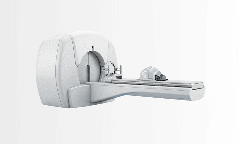
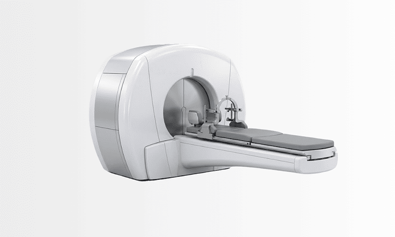
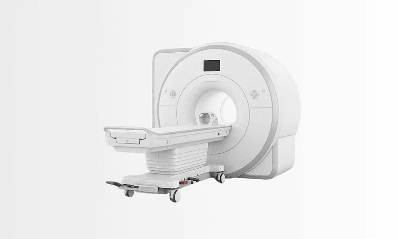
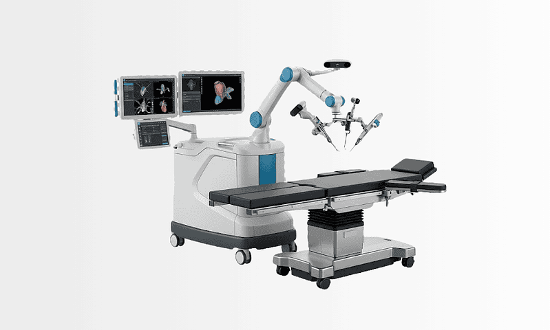
Available at these Hospitals



















What patients say about this unit
★★★★★From 2,400+ verified patient reviews“My wife’s neurosurgery lasted six hours and the family lounge got updates every ninety minutes. Intensive care visits were flexible and the discharge summary was translated for our doctors at home. Professional from start to finish.”
“We flew in terrified after a scan finding; the neurosurgery team re-read the images, ordered one more sequence and downgraded the urgency honestly. When surgery did happen months later it was calm, planned and successful.”
Patient stories from this unit
 Canada Brain Tumor Surgery in Istanbul: Adam’s Journey from Canada
 When Adam, from Canada, was diagnosed with a brain tumor, he chose Acibadem in Istanbul for his surgery, and shared his experience afterwards.
Read the story →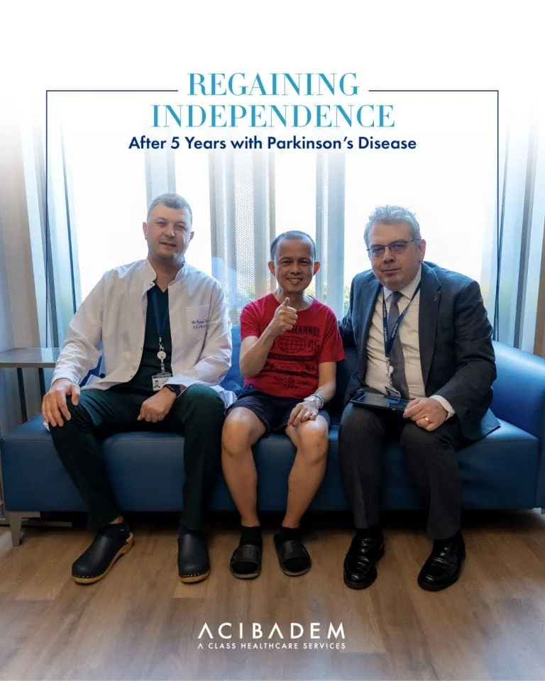 United Kingdom Deep Brain Stimulation for Parkinson’s: Bani’s Journey from London
 A retired IT architect from London, Bani's tremor, stiffness and slowed movement made daily life a struggle, until deep brain stimulation at Acibadem…
Read the story →Comparison guides for this specialty
Speak with our medical team
Share your case and our international patient team will guide you to the right specialist, free of charge.