

Radiation Oncology
IMRT, VMAT and image-guided radiotherapy, SBRT and Gamma Knife radiosurgery, brachytherapy and palliative treatment, planned by radiation oncologists inside a tumour board, on a machine park that includes MR-Linac, CyberKnife and TrueBeam.
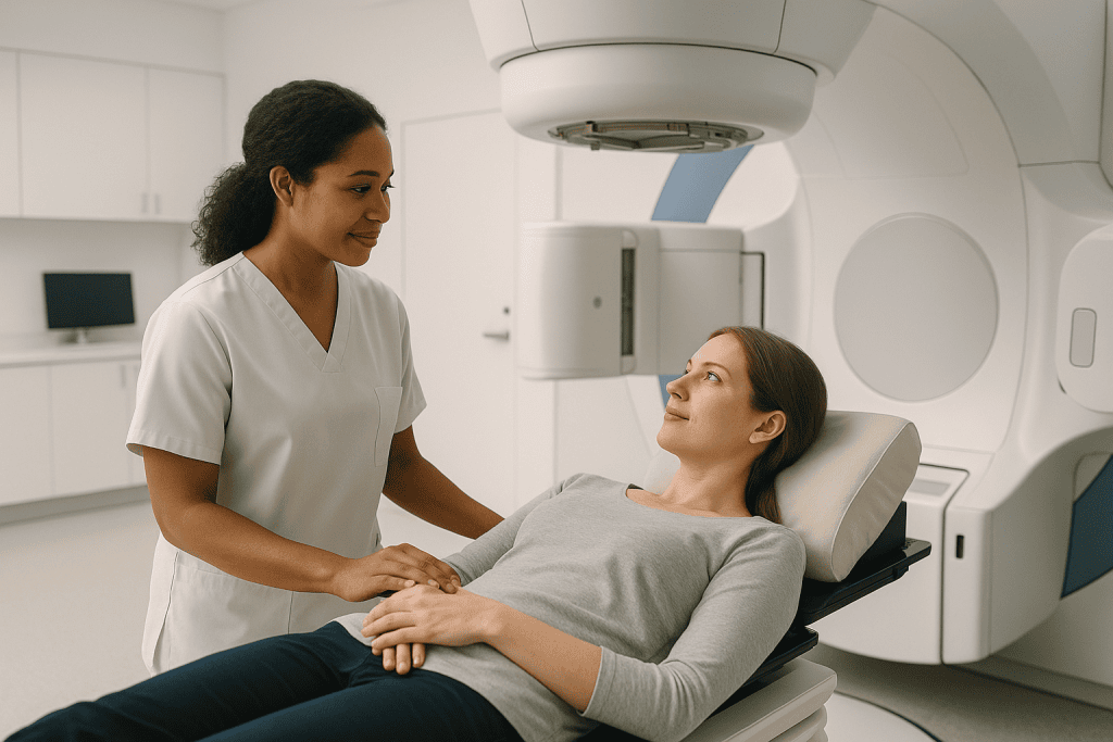
Radiation aimed by a plan, chosen by a board
External beam radiotherapy with IMRT, VMAT and daily image guidance, stereotactic radiosurgery and SBRT, brachytherapy and palliative treatment, one specialty, many instruments, and the instrument always follows the plan.
The machine park
Documented and honestly explained, including the one machine no brochure mentions: the proton centre we do not have, and say so.
Radiation treatments
From the standard daily course to single-session radiosurgery, with the combined-treatment partners that share the calendar.
Cancers where radiation leads
The sites with dedicated sections in the guide, each weighed against surgery and drugs at a tumour board, never in isolation.
The plan is the treatment: the machine only delivers it
Every curative course here passes through a tumour board where the surgeon, the medical oncologist and the radiation oncologist weigh the options together, because for most cancers the real question is not whether radiation is good but in what order, with what, instead of what. The plan is then built by a dosimetrist, signed off by physics independently of the doctors who prescribed it, and aimed again by imaging before every fraction.
And one honest line about machines: Acıbadem does not operate a proton centre. Where a tumour board concludes protons are genuinely preferable, most often for a child with a tumour near the developing brain or spine, the recommendation is a referral to a proton centre, said plainly. A department you can trust about the machines it owns is straightforward about the one it does not.
What we will not do
- Recommend a machine because we own it: the plan and the board choose, and the machine follows.
- Quote a success percentage we could not stand behind for your exact situation.
- Start a curative course before surgery and drug options have been weighed at the same table.
- Soften the ledger, late effects are named before consent, not discovered after.
- Treat palliative as a lesser word: comfort courses are designed with the same care as cure.
Radiation oncologists who lead this work
What actually happens, in order
Records first, flight later
A remote review of your scans, pathology and reports lets the tumour board form a provisional plan before anything is booked, including which schedule family applies, which is the fact that decides everything else about the trip.
The schedule is the trip
Five SBRT sessions, a four-week hypofractionated course and a seven-week chemoradiation course are the difference between ten days and two months in Istanbul. That is knowable in advance, and it is told to you plainly rather than discovered at simulation.
Simulation, then planning days
The CT simulation appointment builds your position, mask and reference marks; the one to two weeks that follow are contouring, dose planning and physics checks on your individual plan. Nothing about the gap is idle: it is simply happening to your scan rather than to you.
The weekday rhythm
Treatment runs Monday to Friday with weekends off, the same short slot each day, interpreter support throughout, and a scheduled weekly review where skin is checked, weight is tracked and the week's questions get their answer.
Leaving with the dose record
The course ends with a written treatment summary and dose record prepared for the oncologist who follows you at home. Radiotherapy's paperwork outlives its appointments: every future doctor deserves to know exactly what was treated, and to what dose.
Six things worth knowing first
Feeling worst after finishing is the described course
Early side effects typically peak in the first one to two weeks after the last session, because tissue keeps expressing damage after the beams stop. It is not treatment failure; it is the curve everyone should be warned about, and it fades as healing overtakes it.
External beam never makes you radioactive
The machine emits radiation only while switched on, exactly as a torch emits light. Between and after sessions there is no source, no residue and no dose to anyone near you: the temporary rules belong only to permanent seed implants and to nuclear medicine therapies.
Side effects are a map of the field
Radiation affects tissue inside the treated volume, so a breast course cannot upset the bowel and a prostate course cannot cause a sore mouth. The one genuinely general effect is fatigue, real, and helped more by light daily activity than by rest.
An easy course is not a failing course
Side effects measure what the beams cross on the way, not what they achieve in the tumour. A gentle course is good fortune rather than weak treatment, and response is judged at the scheduled scan, deliberately set weeks away, so healing tissue is not misread as ambiguity.
Missed days are rescheduled, never dropped
One missed session is added to the end of the course and nothing is lost. Repeated gaps genuinely reduce effectiveness in some cancers, which is why attendance is treated as part of the medicine, and why the final week's sessions matter as much as the first.
Late symptoms are investigated, not assumed
Blood in urine or stool years after pelvic radiotherapy has known radiation causes, and they are diagnoses reached by looking, never by default. "Probably the old radiotherapy" is a conclusion that may only follow a proper examination, not a reason to skip one.
Jump to what you came for
Quick answer
Radiation Oncology is the medical unit that treats cancer and some benign conditions by directing precisely planned radiation at a tumor while protecting as much healthy tissue as possible. At Acibadem in Turkey, this care is delivered through imaging-guided planning and modern radiotherapy techniques by a multidisciplinary team that evaluates each case and organizes treatment accordingly.
What our radiation oncology unit covers
Radiation oncology is the specialty that treats cancer with precisely aimed ionising radiation, radiation therapy in American usage, radiotherapy in British usage, the same treatment under both names. Roughly half of all people treated for cancer receive radiation at some point in their care, sometimes as the main treatment, sometimes around surgery, sometimes purely to relieve symptoms. At Acıbadem International the unit runs across ten hospitals with a machine park that includes linear accelerators, an MR-Linac, Tomotherapy, CyberKnife and two generations of Gamma Knife, and its standing practice is that plans are made the same way everywhere: by a radiation oncologist working inside a tumour board rather than by a machine’s availability.
The work falls into six strands.
- Curative external beam radiotherapyIMRT, VMAT and image-guided treatment for prostate, breast, lung, head and neck, brain, gynaecological, rectal and many other cancers.
- Stereotactic radiosurgery and SBRTvery high doses in one to five sessions for small, well-defined targets in the brain and body.
- Brachytherapyradiation delivered from inside the body, mainly for prostate and gynaecological cancers.
- Radiation with other treatmentschemoradiation, radiation with hormone therapy, and total body irradiation before transplant, planned jointly with the units that own those treatments.
- Palliative radiationshort courses aimed at pain, bleeding or pressure symptoms when cure is not the goal.
- Radiation for skin cancer and selected benign conditionssuperficial treatment where surgery would cost more than it gives, and keloid prevention after excision.
Two boundaries are worth naming at the start, because they are the two most common confusions. Drug treatment for cancer, chemotherapy, immunotherapy, targeted therapy, belongs to medical oncology; this unit works alongside it, often in the same week, but the drugs are not ours. And treatments in which a radioactive substance is swallowed or injected and travels through the body, radioactive iodine, PSMA and receptor-targeted therapies, belong to nuclear medicine. Radiation oncology delivers radiation from outside the body, or from sealed sources placed inside it, for minutes, or permanently with activity that fades over months; nothing we do makes you systemically radioactive, and the one temporary exception, permanent seed implants, is explained in the radioactivity section.
What is radiation therapy (radiotherapy)?
Radiation therapy, radiotherapy in British English, is the use of high-energy ionising radiation to destroy cancer cells in a defined volume of the body while sparing the tissue around it. The radiation itself is usually X-rays of much higher energy than any scan uses, produced by a machine called a linear accelerator; some treatments use gamma rays from a sealed source, or electrons for targets at the skin surface.
People asking what is radiotherapy and people asking what is radiation therapy are asking one question under two flags, and the answer starts from a single fact: it is a local treatment. That fact explains most of what follows: radiation treats the volume the plan draws and nothing else, which is why its side effects are confined to the treated area, why it can be combined with drug treatments that work everywhere, and why it can cure a cancer in one part of the body without touching a cell anywhere else.
What does radiation do to the body?
Radiation damages the DNA inside cells that are dividing. Cancer cells divide constantly and repair themselves poorly, so damage accumulates in them faster than they can fix it; over the days and weeks of a course, the damaged tumour cells die as they attempt to divide. Healthy tissue in the treated area takes some of the same damage, that is what side effects are, but normal cells repair DNA far better than tumour cells do, and the way treatment is scheduled is designed around exactly that difference. Outside the treated volume, the body is not exposed to treatment doses, which is why radiation to a prostate does not cause hair loss and radiation to a breast does not cause nausea.
How does radiation therapy work against cancer, day by day?
How does radiation therapy work in practice? Not by burning the tumour away in one blow, but by arithmetic. The total dose a tumour needs is far more than healthy tissue could tolerate in one sitting, so it is divided into daily doses called fractions. Each fraction injures tumour and normal tissue together; overnight, normal cells repair most of their damage and tumour cells do not. Repeated every weekday, the gap widens until the tumour’s ability to regrow is destroyed. This is also why attending the sessions you find easiest to skip, the ones near the end, when you are tired, matters as much as the first ones, and why the team treats missed days seriously enough to reschedule them.
Is the radiation the same as in an X-ray or CT scan?
It is the same kind of energy at a profoundly different dose and intent: imaging uses the smallest amount of radiation that produces a picture, while radiotherapy delivers a treatment dose shaped to a target, thousands of times higher, on purpose. Comparing the two is like comparing a camera flash to a welding arc, related physics, different instruments, different rules. The daily image-guidance scans taken during a course are at imaging doses and exist to aim the treatment, not to add to it.
Types of radiation therapy
There are three broad types of radiation therapy, defined by where the radiation comes from, outside the body, inside it, or travelling through it, and two intents, curative and palliative, that shape how each is used. Every treatment this unit delivers is one of these types aimed with one of these intents, and naming them early makes the rest of the map easier to read.
External beam radiation, from outside
External beam radiation therapy points shaped beams at the target from a machine that moves around you but never touches you. It is by far the most common form, the daily, Monday-to-Friday treatment most people mean by “radiotherapy”, and it is described in detail in its own section, with the machines themselves in the machine park.
Internal radiation, brachytherapy
Brachytherapy places a sealed radioactive source inside or against the tumour itself, so the dose is highest exactly where the cancer is and falls away within millimetres. It carries its own section, including what it means for the people around you afterwards.
Radionuclide therapy: the border with nuclear medicine
In radionuclide therapy an unsealed radioactive drug is swallowed or infused and finds its target through the bloodstream, radioactive iodine for thyroid cancer is the classic example, PSMA-targeted therapy for prostate cancer the modern one. These treatments are real radiation treatments, but they are planned and delivered by nuclear medicine, not by this unit, and the safety rules around them are different because the radioactivity travels with you for a time. If your plan includes one, that page is the honest place to read about it.
Radical radiotherapy and palliative radiotherapy, two different jobs
Radical radiotherapy means treatment given at full curative dose, alone or with other treatments, accepting more side effects because the goal is to eliminate the cancer. Palliative radiotherapy uses smaller, shorter doses to shrink a tumour that is causing pain, bleeding or pressure, accepting less tumour control because the goal is comfort with the fewest possible hospital visits. The same machines deliver both. Which intent applies to you is a tumour-board decision made before any planning starts, and it is stated openly: a person deserves to know which of the two jobs their treatment is doing. Palliative courses have their own section.
The radiation oncologist (clinical oncologist): the doctor behind the beam
A radiation oncologist is the physician who decides whether radiation should be part of your cancer treatment, prescribes the dose, draws the target on your planning scan, approves the plan, reviews you every week of the course and manages the side effects, in the UK the same doctor is called a clinical oncologist. It is a full medical specialty in its own right: the machines are operated by other professionals, but every clinical decision along the way belongs to this doctor.
What does a radiation oncologist do, visit by visit?
What does a radiation oncologist do in practice? At the first consultation they read your scans, pathology and history and tell you whether radiation has a role at all, sometimes the honest answer is no, or not yet, or not until after surgery. If treatment goes ahead, they define the target volume and the organs to protect, prescribe the total dose and the number of fractions, and sign off the physics plan before the first session. During the course they see you at least weekly, adjust supportive care as side effects appear, and decide on the rare days when treatment should pause. After the course, they follow you until the late picture is clear.
Radiation, drugs and surgery, who decides what
No radiation oncologist decides alone. As standing practice, curative plans at Acıbadem pass through a tumour board where the surgeon, the medical oncologist, the radiation oncologist, the radiologist and the pathologist weigh the options together, because for many cancers the real question is not “is radiation good” but “in what order, with what, instead of what”. Radiation before surgery to shrink, after surgery to secure, instead of surgery to preserve an organ, with chemotherapy to sensitise: each sequence suits different situations, and the board exists so that the recommendation you hear reflects the disease, not the department you happened to enter first.
The team behind every course
A radiation oncology department is a team most patients have never heard of before their first day, and understanding who does what makes it far less intimidating. Four professions stand behind every treatment, and each one checks the others’ work: the layered checking, not any single person, is what makes modern radiotherapy as safe as it is.
The radiation therapist (therapeutic radiographer)
The radiation therapist, therapeutic radiographer in British usage, is the professional you will see most: they run the treatment machine, position you on the couch to the millimetre, take and check the daily guidance images, and deliver the fraction the plan prescribes. What does a radiation therapist do beyond the console? They are also the person who notices day-to-day changes, skin, weight, mood, and flags them to the doctor between your weekly reviews, because they see you every single treatment day and nobody else does.
The dosimetrist: the person who builds your plan
A dosimetrist is the planning specialist who turns the doctor’s prescription into a deliverable treatment: working in the planning system, they shape the beams, set their angles and intensities, and iterate until the tumour volume receives the prescribed dose while each organ around it stays under its limit. What is a dosimetrist to you as a patient? The person you may never meet whose work decides how much of your salivary gland, rectum or heart sits inside the beam, days of skilled work that happen between your planning scan and your first session, which is a large part of why that gap exists.
The medical physicist: the guarantee the machine tells the truth
The medical physicist owns the machines’ honesty: they calibrate each linear accelerator so that the dose it delivers is the dose the plan states, run the quality-assurance measurements on every new plan before a patient is treated with it, and investigate anything that looks even slightly off. Radiotherapy’s safety record rests on this profession’s obsessiveness, and a department is exactly as trustworthy as its physics team is independent, plans are signed off by physics separately from the doctors who prescribe them, which is how it should be everywhere.
The radiation nurse: the side-effect specialist
The radiation nurse manages the human consequences of treatment: skin care as the reaction builds, mouth care and nutrition in head and neck courses, continence and bowel advice in pelvic ones, and the standing answer to “is this normal for week three”. They are the easiest member of the team to reach mid-course, and using them early, at the first sign of a sore mouth or broken skin, not the worst week, is the single behaviour that most changes how hard a course feels.
Planning your course: from CT simulation to the first session
Nothing about radiotherapy is improvised on the day. Between the decision to treat and the first treatment lies a planning process that typically takes one to two weeks, and understanding it removes the most common worry of that gap: that “nothing is happening”. Everything is happening; it is simply happening to your scan rather than to you.
What happens at CT simulation
CT simulation radiation planning starts with a scan that is not a diagnostic test: it is a rehearsal. You lie on a flat couch identical in shape to the treatment couch, in the exact position you will hold for every session, arms up or down, head straight or turned, sometimes with a mask or a moulded cushion made for you there and then. The scanner maps your anatomy in that position, reference marks are set on the skin or mask, and from that day the plan assumes you can be returned to that position to the millimetre. The appointment usually takes thirty to sixty minutes, most of it positioning rather than scanning; for some sites a small amount of contrast, a full or empty bladder, or breath-hold coaching is part of it.
Contouring and radiotherapy treatment planning
After simulation, the radiation oncologist draws, slice by slice through the CT, the tumour volume, the margin around it that accounts for microscopic spread and daily movement, and every organ that must be protected. The dosimetrist then builds the radiotherapy treatment planning solution on that drawing, and the physicist verifies it on the machine before you ever lie under it. Fusing the planning CT with your MRI or PET scans is routine where it sharpens the target, which is one reason bringing complete imaging to the first consultation genuinely changes the quality of what can be drawn.
Fiducial markers and SpaceOAR-style spacers
For targets that move or hide, small helpers are placed before planning. Fiducial markers, gold seeds the size of a grain of rice, placed with a needle under local anaesthetic, make a prostate or a liver tumour visible to the machine’s daily imaging, so the beam follows the target rather than an assumption about it. For prostate treatment a dissolvable hydrogel spacer such as SpaceOAR can be injected between prostate and rectum, pushing the rectal wall a centimetre out of the high-dose region for the months of treatment; whether either is worth it in your case is a planning decision, weighed like any other procedure with its own small risks of bleeding, infection or discomfort, not a default.
The quality checks before day one
Before your first fraction, the plan passes through checks that never involve you: the physics team measures the plan’s actual dose delivery on the machine, the therapists verify the setup instructions, and on day one extra time is taken to image, adjust and confirm before anything is switched on. The first appointment is routinely the longest, often forty-five minutes to an hour against fifteen to twenty for the rest, and knowing that in advance stops it feeling like something has gone wrong. It has not; it is the system proving itself before it treats you.
The radiation mask, tattoos and skin marks
The two objects patients remember most from radiotherapy are the mask and the tattoos, and both exist for the same reason: the plan is only as good as the certainty that today’s position is the planned position. This section is about what they are actually like, because the anticipation is usually worse than the object.
The radiotherapy mask, honestly described
For treatment of the head, brain or neck, a radiation mask: a thermoplastic mesh warmed in water, laid over your face and shoulders, and moulded to you in about ten minutes as it cools, holds your head still to within a millimetre or two, carries the reference marks so your face does not have to, and lets the same position be rebuilt every day. It clips to the couch, it feels snug rather than crushing, you can breathe and swallow normally in it, and each session in it lasts minutes rather than hours.
Claustrophobia and the mask, what actually helps
A meaningful minority of people find the mask genuinely hard, and departments have real answers rather than pep talks: eye holes can often be cut open, the fit can be adjusted, music and a hand signal agreed in advance restore a sense of control, and a short-acting medicine before sessions can be arranged with the team for the people who need it. Telling the therapists at the mask-making appointment, not silently enduring until week three, is what makes those adjustments possible from day one.
Radiation tattoos and the no-tattoo alternative
A radiation tattoo is a permanent ink dot the size of a freckle, usually three of them, one central and one on each side, placed at the simulation appointment so your alignment survives showers, weeks and skin creams. Many people barely notice them afterwards; some genuinely mind carrying a permanent mark of treatment, and that feeling deserves to be said out loud at simulation rather than swallowed. Surface guided radiation therapy (SGRT), camera systems that track the skin surface itself, lets many treatments, breast treatment especially, run with no permanent marks at all, and where it is available and suitable the team will say so.
What a treatment day is actually like
A course of external beam radiotherapy is a rhythm: the same machine, the same therapists, the same few minutes, every weekday for one to eight weeks depending on the prescription. Most people are surprised by two things, how short the sessions are, and how normal life continues around them. What follows is the honest shape of a treatment day; the separate sections on side effects cover how the weeks feel as they accumulate.
How long does radiation take?
How long does radiation take, session by session and course by course?
- In the room: usually 15–20 minutes, of which the beam itself runs for only 2–5. The rest is positioning and imaging.
- First session: the longest, often 45–60 minutes, because everything is verified before treatment starts.
- Per week: almost always Monday to Friday, with weekends off for normal tissue to repair.
- Whole course: anywhere from a single session for pain relief to around eight weeks for the longest curative schedules: the families of schedules are explained under external beam radiotherapy.
Does radiation hurt?
Does radiation hurt while it is being given? No: you feel nothing during the beam itself, exactly as you feel nothing during an X-ray; there is no heat, no sting, and the machine never touches you. What people actually experience in the room is stillness, the quiet hum and repositioning of the gantry, and the therapists’ voice over the intercom. Discomfort in radiotherapy comes later and cumulatively: the skin, mouth or bowel reactions described under side effectsnever from the beam itself, though holding a position can be its own effort when bones already hurt, and the therapists adjust supports for exactly that.
Alone in the room, never unwatched
For the beam to run, the therapists step out: you are the only person in the room, which unsettles some people the first time. You are on camera and intercom the entire time, a raised hand pauses treatment within seconds, and the therapists can be back at the couch in moments. Many departments let you bring your own music; ask at the first session. The door is heavy and the walls are thick for physics reasons that have nothing to do with how much radiation is in the room between beams: none.
The weekly review
Once a week, usually attached to a treatment day, you see the radiation oncologist, often together with the radiation nurse, for a scheduled review: skin checked, weight tracked, side effects graded, supportive care adjusted. This is the built-in moment for every question the week has generated, writing them down as they occur to you and bringing the list beats trying to remember at the door.
External beam radiation therapy, technique by technique
External beam radiation therapy (EBRT) is the workhorse of the specialty: shaped photon beams from a linear accelerator, entering from multiple angles that each cross healthy tissue at a tolerable dose and add up to a treatment dose only where they intersect. The techniques in this section are stages of one idea, ever finer control over where the dose lands, and most modern treatments use several of them at once.
From 3D conformal to IMRT
Three-dimensional conformal radiotherapy shapes each beam to the target’s outline as seen from that beam’s angle; it remains a sound technique for simple geometries. Intensity modulated radiation therapy, IMRT, goes further: each beam is divided into thousands of beamlets whose intensities are individually tuned, so the dose can wrap around a curved structure, treating a horseshoe-shaped target while the spinal cord in its middle stays under limit. IMRT radiation is the reason head and neck treatment can now routinely spare a salivary gland that once sat helplessly in the field.
VMAT: the arc version
VMAT radiation (volumetric modulated arc therapy) delivers IMRT while the machine sweeps around you in continuous arcs, modulating shape, intensity and speed as it moves. The plans are as precise as fixed-field IMRT and considerably faster to deliver, often two to four minutes of beam, which matters for comfort, for breath-hold techniques, and for holding perfectly still; where it suits the plan, it is the technique modern departments reach for first.
IGRT, aiming before every fraction
Image guided radiation therapy is the discipline of imaging you on the treatment couch, cone-beam CT, planar X-rays or surface tracking, immediately before each fraction, and shifting the couch until today’s anatomy matches the plan’s. IGRT is what turns tight margins from a gamble into an engineering statement, and it is treated as part of the treatment itself rather than an upgrade. It is also the safety net that catches the slow changes, weight loss, tumour shrinkage, that can quietly invalidate a plan, triggering a re-scan and re-plan when the pictures stop matching.
Hypofractionated radiation: the schedule families
Hypofractionated radiation means fewer, larger daily doses, and it is where treatment schedules have moved wherever the evidence supports it. The families of schedules, set out once here, and only pointed to from the rest of the guide, are:
- Conventional fractionation: small daily doses over roughly six to eight weeks, still standard for some head and neck, gynaecological and other sites.
- Moderate hypofractionation: roughly three to four weeks, now a standard for many breast and prostate treatments.
- Ultra-hypofractionation / SBRT-style schedules: one to five larger sessions for selected small targets, described under stereotactic treatment.
- Palliative schedules: a single session or five, chosen for the fewest visits that relieve the symptom.
Which family your prescription belongs to is a clinical decision built on trial evidence for your exact situation, shorter is not a favour and longer is not a punishment, and the honest sentence is that the right schedule is the one tested for your disease, stage and anatomy.
The boost, extra dose where recurrence starts
A radiation boost is an additional, smaller-volume dose to the highest-risk part of the target: the tumour bed after breast surgery, the visible tumour inside a larger treated region, delivered as extra sessions at the end of a course or woven into it. If your schedule ends with “plus a boost”, that is what it means: the same machine, a smaller field, the finishing nail.
The machine park, honestly explained
Patients are shown machine names as if they were treatments, and the honest starting point is this: the plan, the imaging and the team matter more than the badge on the gantry, and a good department chooses the machine to fit the plan rather than selling the machine it has. Acıbadem’s radiation oncology units operate the platforms in this section across its hospitals; each name links to a page describing it properly.
What is a linear accelerator?
What is a linear accelerator? The general-purpose radiotherapy machine: it accelerates electrons almost to light speed, converts their energy into a high-energy X-ray beam, shapes that beam with a multileaf collimator, dozens of tungsten leaves that move independently, and delivers IMRT, VMAT, IGRT and stereotactic treatments as the plan requires. When people picture “the radiotherapy machine”, the large rotating gantry with the flat couch, they are picturing a linear accelerator; the TrueBeam platform in use at Acıbadem is a modern example built for image-guided and stereotactic work.
MR-Linac, radiotherapy that watches soft tissue live
The MR-Linac combines a linear accelerator with a diagnostic-quality MRI scanner in one machine, imaging soft tissue in real time while the beam runs. MRI guided radiation therapy earns its keep where soft-tissue targets move and CT sees them poorly, pancreas, liver, prostate, targets near bowel, and it enables daily adaptive radiation therapy: reshaping the plan to that day’s anatomy while you lie on the couch, rather than treating today’s body with last week’s plan.
Ethos and adaptive radiation therapy
Adaptive radiation therapy is the same idea, re-planning to match the anatomy of the day, built on CT imaging with artificial-intelligence-assisted contouring: the Ethos platform generates an adapted plan of the day in minutes, with the physician and physicist approving it while you remain in position. Where daily anatomy changes meaningfully, a bladder that fills, bowel that drifts into yesterday’s safe corridor, adaptation is the difference between the plan you were prescribed and the plan you actually receive.
Tomotherapy: CT geometry, helical delivery
Tomotherapy delivers IMRT helically, slice by slice, from a machine built on CT geometry: the beam spirals around you as the couch moves through the bore. Its strengths are very long or complex volumes: craniospinal treatment, total marrow irradiation, targets that would force awkward field junctions on a conventional gantry, where a single continuous helix treats the whole volume without seams.
CyberKnife: the robotic radiosurgery specialist
CyberKnife mounts a compact linear accelerator on a robotic arm that delivers narrow beams from hundreds of angles, tracking the target, including targets that move with breathing, via implanted markers, and correcting in real time. It is a dedicated instrument for stereotactic radiosurgery and SBRT: small, well-defined tumours at the highest precision, in one to five sessions, frameless.
Gamma Knife: Esprit and Perfexion
The Gamma Knife: Acıbadem operates both the Perfexion and the newer Espritfocuses nearly two hundred fine cobalt beams on a single point, achieving sub-millimetre precision for targets inside the skull only. It treats brain metastases, acoustic neuromas, meningiomas, arteriovenous malformations and trigeminal neuralgia, usually in a single session. Whether a particular brain lesion is best served by radiosurgery, microsurgery or watching is a joint decision with neurosurgery, whose page walks through that choice honestly; the machine itself lives in this unit’s world, the decision lives in both.
Stereotactic radiosurgery (SRS) and SBRT, one to five sessions, surgical precision
Stereotactic radiosurgery is radiation used like a scalpel: a very high dose delivered with sub-millimetre accuracy to a small, sharply defined target, in a single session or a handful, with steep fall-off that spares what sits a few millimetres away. Despite the name, nothing is cut, “surgery” describes the precision and the one-off nature, not the method. Inside the skull the approach is called SRS; in the rest of the body it is SBRT, and the two share physics, machines and philosophy.
SBRT, stereotactic body radiation therapy
Stereotactic body radiation therapy (SBRT) treats small, well-defined tumours in the body, early lung cancers, liver and pancreatic tumours, prostate cancer, kidney tumours, isolated metastases and spinal targets, in typically one to five sessions instead of weeks. SBRT radiation demands everything the planning sections describe at once: rigid immobilisation, management of breathing motion by tracking, gating or breath-hold, image guidance before and during every fraction, and physics margins measured in millimetres. Its rise is one of the genuine changes in modern oncology: for a medically inoperable early lung cancer, for selected patients with a limited number of metastases, and for prostate treatment in five visits, it has turned “weeks of radiotherapy” into “days”.
SABR: the same treatment under its British name
SABR, stereotactic ablative radiotherapy, is the UK’s name for exactly what SBRT names in American usage; if your letters say SABR or stereotactic radiotherapy and this unit’s plan says SBRT, no translation is needed beyond that sentence. The schedules, machines and evidence base are the same, which also means UK guidance about SABR eligibility reads across directly to a treatment planned here.
SBRT for lung tumours: the flagship case
SBRT lung cancer treatment is the clearest example of what the technique is for: a small peripheral lung tumour in a person whose breathing reserve or other illness makes surgery unwise can be treated in three to five outpatient sessions, breathing managed rather than suppressed, with no incision and no anaesthetic. The lung section carries the decision context, how it is weighed against surgery, and by whom.
SRS for brain metastases
For a limited number of brain metastases, SRS in a single session, on Gamma Knife, CyberKnife or a stereotactic linear accelerator, has largely replaced whole-brain treatment as the first radiation move, precisely because it treats the visible lesions and leaves the rest of the brain out of the field. When lesions are many or diffuse, whole brain radiation therapy still has a role, and the trade-offs are laid out plainly in the brain section. For benign targets, acoustic neuroma, meningioma, trigeminal neuralgia, the decision between radiosurgery and microsurgery is walked through on the neurosurgery page.
Brachytherapy, radiation from the inside
Brachytherapy places a sealed radioactive source inside the tumour or against it, so the highest dose in the entire plan sits exactly where the cancer is and falls away within millimetres: the inverse of external beam, where every dose must first cross healthy tissue to arrive. It is a procedure as much as a treatment: sources travel through needles, catheters or applicators placed under anaesthetic or sedation, guided by ultrasound, CT or MRI, and the physics is planned around the placement in real time.
HDR brachytherapy, minutes, then nothing remains
HDR brachytherapy (high dose rate) drives a single tiny source, attached to a wire, through pre-placed applicators under computer control: it dwells at planned positions for seconds to minutes, delivers the fraction, and withdraws completely, leaving nothing in the body; what that means for the people around you is stated with every other radioactivity rule in one section. HDR is the workhorse for gynaecological treatment and one of the standard options for prostate treatment, either alone or as a boost added to external beam.
LDR seeds, brachytherapy for prostate cancer
LDR (low dose rate) brachytherapy for prostate cancer implants dozens of rice-grain-sized radioactive seeds permanently, in a single anaesthetic, delivering their dose gradually over months as the isotope decays. Brachytherapy for prostate cancer in this form suits selected smaller, lower-risk cancers, and it is the one treatment in this unit that carries temporary precautions afterwards, stated once, with every other radioactivity rule, in the radioactivity section.
Brachytherapy for cervical cancer
Brachytherapy for cervical cancer is not an optional extra: in curative treatment of cervical cancer it is the component that delivers the tumour-killing dose external beam alone cannot reach safely, and international guidance treats a course without it as incomplete. Applicators are placed under anaesthetic, imaging, increasingly MRI, verifies their position, and a small number of HDR fractions complete the plan alongside pelvic external beam and chemotherapy. Gynaecological cancers themselves are the territory of gynaecology and obstetrics; the radiation component is planned here, jointly.
Safety around others, answered once
The question every brachytherapy patient asks, can I hold my grandchild, has a precise answer that depends on which form you had, and it is answered together with every other radioactivity question in one section, so that no two versions of the answer can drift apart.
Proton therapy (proton beam therapy), and an honest note on access
Proton therapy replaces X-ray photons with accelerated protons, whose physics offer one genuine gift: a proton slows and stops, dumping most of its energy at a chosen depth, the Bragg peak, with almost nothing beyond it. Proton beam therapy therefore puts a hard stop behind the target, which photons cannot, and that makes it genuinely valuable in a specific set of situations rather than a general upgrade.
Proton therapy vs radiation with photons
Proton therapy vs radiation of the conventional kind is not a contest photons are losing: for most adult cancers, modern IMRT and VMAT achieve clinically equivalent tumour doses with organ sparing good enough that trials keep failing to show protons add cure. The exit-dose advantage matters most where every spared gray counts for decades or where critical structures sit directly behind the target: children and young adults, tumours of the skull base and spine, re-irradiation of previously treated tissue, and selected head and neck or eye tumours. For those situations protons are a real consideration; for the rest, the machine park treats to the same standard of evidence.
Proton therapy cost and access, stated plainly
Proton therapy cost is the bluntest fact about it, several times that of photon treatment wherever it is offered, and Acıbadem does not operate a proton centre; that is stated here because a unit that explains every machine it has should also say which one it has not. Where a tumour board concludes that protons are genuinely preferable for a specific case, most often a child with a tumour near the developing brain or spine, the honest recommendation is a referral to a proton centre, and that is the recommendation that gets made. A department you can trust about the machines it owns is one that is straightforward about the machines it does not.
Chemo vs radiation, different jobs, often the same plan
Chemo vs radiation is the wrong fight to pick, because they are not competitors: chemotherapy is a systemic treatment that travels everywhere blood goes, radiation is a local treatment confined to the volume in the plan, and modern cancer care constantly combines the two precisely because each covers the other’s blind side. The real questions are sequence and purpose, and they have honest answers.
Radiotherapy vs chemotherapy, when each leads
Radiotherapy vs chemotherapy is settled by the disease’s geography. A cancer that is localised and needs local control leads with surgery or radiation; a cancer that is widespread, or carries high risk of unseen spread, needs drugs that travel. That is why early prostate or larynx cancer may be treated with radiation alone, why leukaemias are treated with drugs and almost never with beams, and why many solid tumours in between receive both, drugs for the invisible disease, radiation for the visible one. Neither modality is “stronger”; they answer different questions.
Chemoradiation, given together, on purpose
Chemoradiation means chemotherapy delivered during the radiation course, usually lighter doses than stand-alone chemotherapy, because certain drugs make tumour cells measurably more vulnerable to each fraction. It is the established curative standard for several cancers treated without surgery, cervix and many head and neck and oesophageal cancers among them, and it is deliberately harder than either treatment alone: the side effects of both arrive in the same weeks. When a plan proposes it, that is not enthusiasm; it is trial evidence that the combination cures where either alone does not. The drug half is prescribed and supervised by medical oncology, the radiation half here, on one shared calendar.
Hormone therapy with radiation
For prostate cancer specifically, androgen deprivation, hormone therapy, is often prescribed before, during and after radiation for intermediate and higher-risk disease, because lowering testosterone makes the radiation measurably more effective and treats microscopic disease beyond the field. How long it continues is a risk-based decision made by the treating doctors; its side effects are real and deserve their own conversation with whoever prescribes it. The prostate section carries the radiation half of that story.
Radiation side effects (radiotherapy side effects): the honest map
Radiation therapy side effects follow rules, and knowing the rules in advance converts most frightening surprises into expected events. Radiation side effects are local, cumulative and largely predictable from the treated area, radiotherapy side effects, under the UK name, follow exactly the same rules: this section owns the timing, the skin and late-effects sections own their specifics, and each body-site section names what its patients actually meet.
Why side effects stay local
Radiation only affects tissue inside the treated volume, so the side-effect list is a map of the field: a breast course cannot cause bowel trouble, a prostate course cannot cause a sore mouth, and hair falls only where beams pass. The two genuine exceptions are fatigue, which is general, and the rare effects of the small scattered dose outside the field, which belong to the late-effects discussion. If a symptom appears during treatment in a part of the body the plan does not touch, it deserves assessment on its own merits rather than automatic blame on the radiation.
The timing rules, early effects, the peak, and the fade
Early side effects build gradually: the first week usually feels like nothing, effects appear from the second or third week as damaged surface cells fail to renew, and, the single most useful fact in this section, they typically peak in the first one to two weeks after the last session, because the tissue keeps expressing damage after the beams stop. Feeling worst ten days after finishing is not treatment failure; it is the described course. Most early effects then settle over the following weeks as surfaces heal. Late effects, arriving months to years afterwards, follow different biology and are treated separately under their own names.
Radiation fatigue
Radiation fatigue is the one genuinely general side effect: a specific, heavy-limbed tiredness that accumulates over the weeks, differs from sleepiness, and is not proportional to how “small” the treated area is. It is real, it is not weakness, and the evidence-backed response is unintuitive, regular light activity, a short daily walk more than extra bed rest, protected sleep, and honest workload adjustment rather than pushing through at full load. It typically deepens toward the course’s end, plateaus, and lifts over the weeks after finishing, more slowly than most people expect.
Does radiation cause hair loss?
Does radiation cause hair loss? Only where the beams pass: this is the local rule at its most visible. Scalp hair is lost only when the head itself is treated; chest, armpit or pubic hair goes only if those areas sit in a field; and a prostate, breast or lung course does not touch the hair on your head, a point worth stating because chemotherapy’s pattern is the opposite and the two treatments are so often given in the same season of life. Where treated-area hair does fall, it usually begins two to three weeks in, and regrowth after moderate doses arrives over months, sometimes changed in texture, and after very high doses to the scalp, sometimes incomplete. What will happen in your plan’s geography is a question the team can answer from the dose map before the first session.
Skin during radiation, dermatitis, burns and daily care
The skin is the one organ that sits in the path of almost every external beam, and its reaction is the side effect most people can see coming and want rules for. Modern techniques have made severe reactions far less common than the stories people arrive with, but the skin inside a treated area still lives through the course, and it does best with boring, consistent care rather than heroics.
Radiation dermatitis, what the reaction actually looks like
Radiation dermatitis is the medical name for the treated skin’s reaction: typically nothing in week one, then pinkness and dryness like mild sunburn, sometimes deepening to darker colour, itching and peeling as weeks accumulate, worst, per the timing rules in the side-effects sectionshortly after the course ends, then healing over the following weeks. Skin folds, the armpit in breast treatment, the groin in pelvic work and the neck in head and neck courses react most, because folds add friction and moisture to the dose. Longer-term, treated skin can stay slightly darker, smoother or drier than its neighbours, and it remains more sun-sensitive permanently: a high-factor sunscreen habit for the treated patch is a lifelong souvenir worth keeping.
Radiation burns: the honest name for the severe end
Radiation burns, moist desquamation, in the clinic’s language, are the severe end of the same spectrum: skin that breaks, weeps and exposes raw surface, most often in folds and most often in the final weeks of long courses or just after. The team grades skin at every weekly review precisely to see this coming; broken skin gets specific dressings and care from the radiation nurse, and it heals, skin regenerates impressively once the course ends. What turns a manageable reaction into a miserable one is usually silence: a patch that broke on Friday and was endured until Thursday. Showing the team early is the entire strategy.
Cream for radiation burns, and what actually helps day to day
Cream for radiation burns is the most-searched phrase in this territory, and the honest answer has two halves. Day to day: wash the treated area gently with lukewarm water and mild unperfumed soap, pat dry, keep an unperfumed moisturiser on unbroken skin from the start of the course, wear soft loose layers, and shield the area from sun, heat packs, ice and adhesive tape. Just as important: nothing new, creams, oils, home remedies, goes on before the treatment team has seen it, partly because broken skin needs dressings rather than creams, and partly because the right product depends on the state of your skin that week; the radiation nurse reviews it and adjusts the plan as the reaction evolves.
Late effects, named plainly
Months to years after a course, a different family of changes can appear in the treated area, not a return of the acute reaction but slower remodelling of tissue: small vessels narrow, flexible tissue stiffens, glands that stopped may not restart. Most people never meet most of these. They are named here individually and honestly, because people meet the words in follow-up letters and search results, and an unexplained name is more frightening than an explained one, and because a unit that delivers the treatment owes its patients the whole ledger, not the bright half.
Radiation fibrosis
Radiation fibrosis is the umbrella process behind several late effects: treated tissue gradually replaces some of its elasticity with firmer scar-like tissue, which can mean a firmer area of breast, a stiffer patch of neck, reduced flexibility of a joint capsule inside a field, or, in the lung, a stable scarred patch visible on every future scan. It develops slowly, tends then to stabilise, and cannot be reversed, which is exactly why modern planning spends so much effort keeping dose away from tissue that does not need it; once established, care is about maintaining movement and function, where physiotherapy in the rehabilitation unit genuinely earns its place.
Radiation proctitis
Radiation proctitis is inflammation and vessel fragility of the rectal lining after pelvic courses, prostate, cervix, rectum, showing itself as urgency, mucus or intermittent bleeding, occasionally beginning years after treatment. The bleeding pattern is characteristically small and recurrent rather than dramatic, but bleeding is never assumed to be proctitis by default: it is examined, because the rectum can develop unrelated disease with the same symptom, and the diagnosis is made by looking, in the gastroenterology unit. Established proctitis has real treatments, from local anti-inflammatory measures to endoscopic treatment of the fragile vessels, and rectum-sparing techniques and spacers exist precisely to make it rarer.
Radiation cystitis
Radiation cystitis is the bladder’s version: a bladder wall inside a pelvic field can become inflamed and later fragile, producing frequency, urgency, discomfort or visible blood in urine, months to years after treatment. Blood in urine after pelvic radiation is investigated properly every time, cystoscopy in the urology unitbecause “probably the old radiotherapy” is a conclusion that may only be reached after new disease is excluded, never a reason not to look. Management ranges from bladder-calming medicines to endoscopic treatment of bleeding points in the harder cases.
Radiation pneumonitis
Radiation pneumonitis is an inflammatory lung reaction that can follow chest courses, lung, oesophagus, some breast and lymphoma fields, typically arriving weeks to a few months after finishing, as a dry cough, breathlessness on effort or low-grade fever. It is treatable, usually with a supervised course of corticosteroids prescribed by the treating doctors, and it matters because its timing tricks people: appearing after treatment has ended, it is easily mistaken for infection or for the cancer, and it is diagnosed by putting the scan, the field map and the timeline side by side with the pulmonology unit. Over time a treated patch can leave the stable scar described under fibrosis: a finding future radiologists should know the treatment history to read correctly.
Radiation esophagitis and strictures
Radiation esophagitis, oesophagitis in British spelling, is soreness of the swallowing tube when it sits within a chest field, and it is the one early effect named in this section, because it belongs beside its late consequence, the stricture: heartburn-like pain and painful swallowing in the mid-course weeks, managed with food texture changes, liquid analgesia and attention to hydration until it settles after the course. Uncommonly and much later, a healed segment can narrow into a stricture that makes solids stick; strictures are mechanical problems with a mechanical answer, endoscopic dilation in the gastroenterology unitnot something to live around silently.
Radiation recall
Radiation recall is the strangest entry: weeks to years after a course, certain drugs, several chemotherapies and some newer agents, can wake the old field, producing a sharply bordered skin reaction exactly where treatment once was, like a photograph re-developing. It looks alarming and means something specific rather than sinister: the combination of that drug and the treated tissue. It settles with drug adjustment and skin care, decided by the prescribing team together with the radiation oncologist, worth knowing about mainly so that a rectangle of red skin years later is recognised for what it is rather than feared.
Second cancers, does radiation cause tumors?
Does radiation cause tumors of its own? Rarely, yes: a radiation-induced second cancer can arise in or at the edge of a treated area, typically a decade or more later, and pretending otherwise would be the kind of reassurance this unit does not sell. The risk is small, it is smallest with the conformal techniques that keep unnecessary tissue out of the field, and for people treated for an existing cancer it is overwhelmingly outweighed by the treatment’s benefit: that arithmetic is precisely why the tumour board treats radiation as a serious decision rather than a default, why children’s plans are drawn with decades in mind, and why lifelong participation in normal screening matters after treatment. Stating this plainly is part of consent, and any account of radiotherapy that omits it is incomplete.
Eating, working and daily life during a course
Most people continue a recognisable version of normal life through external beam treatment, and the honest picture is neither “carry on as if nothing is happening” nor “clear your calendar for two months”: it is a rhythm with a daily appointment in it, side effects that depend on geography, and energy that needs budgeting in the later weeks.
Foods to avoid during radiation, and the diet that actually helps
Foods to avoid during radiation depend on where the beams pass, not on radiation itself: pelvic and abdominal courses often go easier on a temporarily gentler, lower-insoluble-fibre pattern if the bowel is reacting; head and neck and oesophageal courses shift toward soft, moist, non-acidic, non-spicy food as the lining gets sore; breast, brain and limb treatments need no food rules at all. Two rules hold everywhere: weight loss during treatment is never ignored, in mask-based treatment it can literally change the fit, and a re-plan is sometimes needed, and supplements or restrictive “anti-cancer” diets started mid-course are shown to the team first, because extreme regimens during treatment cost muscle exactly when it is needed. The clinical nutrition unit is part of the department’s routine, not a luxury referral.
Working, driving and exercise
Many people work through treatment, especially in the first half of a course, and many adjust to part-time or lighter duties in the later weeks as fatigue accumulates, both are normal, and the schedule’s predictability (the same fifteen-minute slot each weekday) makes planning around it easier than most jobs fear. Driving is generally unaffected by external beam treatment itself, with the honest exceptions of brain treatment, seizure history and heavy sedation for masks, where the rules are individual and belong to the doctor’s review. Regular light exercise is not merely permitted but is the best-evidenced remedy for radiation fatigue; the practical ceiling is chlorine and friction, swimming pools are usually paused while treated skin is reacting, and gym gear that rubs the field waits until the skin settles.
Are you radioactive after radiation therapy?
Radioactive after radiation therapy: the fear those two words carry separates families from patients at exactly the moment closeness matters, so the answer deserves to be stated with complete clarity, form by form. It is also the section where every other part of the page sends its version of the question, so the answer exists exactly once.
External beam, no, at any point
External beam radiotherapy leaves nothing radioactive in you: the machine emits radiation only while switched on, exactly as a torch emits light, and when the beam stops there is no source, no residue and no dose to anyone near you. From the first fraction to the last, you can hold children, share a bed, sit beside a pregnant colleague and hug whoever you like, without a single precaution. Any memory of “keeping distance after radiotherapy” someone offers you comes from the other two forms, or from folklore.
Implants and radionuclides: the two real exceptions
Two treatments genuinely carry temporary rules. Permanent LDR seed implants keep emitting a small, steadily fading dose for some weeks: the team gives concrete time-and-distance guidance, mostly limiting prolonged close contact with pregnant women and small children for a defined period, and a card to carry because the seeds can trigger sensitive security detectors. And unsealed radionuclide therapies, radioactive iodine and its relatives, given in nuclear medicinemake you measurably radioactive for days, with specific household rules that unit provides in writing. HDR brachytherapy, by contrast, leaves nothing behind between or after sessions; its rules end when the source is withdrawn.
Pregnancy in the household, and pregnancy in the patient
A pregnant partner, daughter or colleague needs no protection from someone receiving external beam treatment, and after HDR brachytherapy none either; only the seed-implant and radionuclide rules just described mention pregnancy at all, and then only about prolonged closeness for a limited time. Pregnancy in the patient is an entirely different matter: radiotherapy during pregnancy is a highly specialised situation planned case by case, and the department asks about the possibility of pregnancy before planning scans and treatment precisely because it changes decisions. Fertility after pelvic treatment is a real consideration in both sexes, options for preserving it are discussed before a curative pelvic course, in time to act on them.
Radiation for prostate cancer
Radiation for prostate cancer is one of the two curative paths for localised disease, the other being surgery, and for many men the genuinely hard part is not the treatment but the choice, because both paths cure the cancers they are suited to and differ mainly in how their side effects are distributed. This section explains the radiation path; the surgical one belongs to the urology unit, and the comparison belongs to both, made honestly with the same numbers on the table.
The radiation options for prostate cancer
External beam treatment for prostate cancer now spans the three schedule families set out under external beam radiotherapymoderate hypofractionation as a common standard, SBRT for suitable cases, and longer conventional schedules where anatomy or risk argues for them, all delivered with fiducial-marker image guidance, as described in the planning section. Brachytherapy adds two more: LDR seeds alone for selected lower-risk disease, or an HDR boost combined with external beam for higher-risk cancers. Hormone therapy joins the plan for intermediate and higher risk, as described under combined treatment. Which options genuinely apply is a function of risk group, prostate size, urinary function and MRI anatomy, which is why two men at the next-door machines can be on entirely different schedules, both correct.
How many radiation treatments for prostate cancer?
How many radiation treatments for prostate cancer a plan prescribes follows the schedule families that the external beam section owns: five sessions in SBRT form, roughly four weeks in moderate hypofractionation, seven to eight weeks conventionally, with brachytherapy compressing its contribution into one or two procedures. The count is chosen from trial evidence for your risk group and anatomy, not from convenience, though when two schedules are genuinely equivalent for a case, the shorter one is a legitimate thing to prefer and to ask for, and for a man flying in from abroad it often decides the plan’s practicality.
Prostate radiation side effects
Prostate radiation side effects follow the local rule: during the course and shortly after, urinary frequency, urgency, weaker stream and bowel irritability from the rectum’s front wall sharing the neighbourhood; these build per the timing rules and settle for most men over the weeks after finishing. The honest longer view: a minority carry ongoing urinary or bowel changes: the proctitis and cystitis entries name the named ones, and erectile function tends to decline gradually over the years after treatment rather than abruptly, a curve steepened when hormone therapy is part of the plan. Spacers and image guidance exist to shrink the bowel side of this ledger, and continence is affected differently than after surgery, which is exactly the kind of difference the next heading is for.
Surgery vs radiation for prostate cancer
Surgery vs radiation for prostate cancer has a fair summary: cure rates for comparable risk groups are similar, and the choice is really between two side-effect profiles and two relationships with time. Surgery concentrates its costs early: an operation, catheter days, an incontinence risk that improves over months, immediate erectile change with gradual recovery, and gives the pathology report’s certainty; radiation spreads its costs, no operation, minimal incontinence risk, bowel exposure instead, erectile decline arriving gradually, and keeps surgery available afterwards only with added difficulty, while salvage radiation after surgery is well-trodden. Age, urinary symptoms, bowel disease, anaesthetic risk and personal weighting of those profiles decide it; what the decision deserves is both specialists, the same scans, and no salesmanship from either room.
Radiation for breast cancer
Radiation for breast cancer most often follows breast-conserving surgery, where it does a precisely stated job: treating the microscopic disease a lumpectomy cannot see, so that keeping the breast carries the same survival as removing it: the pairing that makes breast conservation legitimate at all. The surgical decisions themselves, including mastectomy, reconstruction and single-dose intraoperative radiotherapy where it applies, live with the breast health unit; this section is about what the radiation course itself involves.
Radiation after lumpectomy: the standard course
Radiation after lumpectomy today usually means a moderately hypofractionated course of roughly three to four weeks: the schedule families apply, treating the whole breast, sometimes with a boost to the tumour bed for higher-risk findings, and sometimes including nearby lymph node regions when the pathology argues for it. Partial-breast and five-session schedules exist for carefully selected cases, and radiation after mastectomy has its own indications when the tumour or nodes carried higher risk. Which shape applies is set by the pathology report and the tumour board, which is why two women with “breast radiotherapy” can attend for different numbers of weeks and both be treated by the book.
Deep inspiration breath hold, protecting the heart
For left-sided breast treatment, deep inspiration breath hold (DIBH) is the elegant trick that moves the heart out of the conversation: a held deep breath inflates the lungs and swings the heart down and away from the chest wall, the beam runs only while the breath is held and the surface-tracking cameras confirm it, and the heart’s dose falls substantially for the price of practising a breathing pattern. It is standard technique, coached at simulation, and one of the clearest examples of how much of modern radiotherapy is geometry rather than force.
Breast radiation side effects
Breast radiation side effects concentrate on the skin and the treated breast itself: the dermatitis spectrum with the armpit fold as its focal point, breast swelling and tenderness during and shortly after the course, then, over months, the possibility of a firmer, slightly smaller or slightly darker treated breast as the fibrosis entry describes. Fatigue follows its usual arc. When lymph node regions are treated, arm swelling becomes a long-term consideration alongside surgery’s contribution; the breast health unit owns the lymphoedema story in full, including the monitoring and therapy that make it manageable. Lung and heart doses in modern breast plans are kept low by design, DIBH being one of the tools, and the rare late lung patch belongs to the pneumonitis entry.
Lung cancer radiation: SBRT to chemoradiation
Lung cancer radiation, radiotherapy for lung cancer, in UK usage, spans the specialty’s whole range: curative SBRT in a handful of sessions for small early tumours, full-dose chemoradiation for locally advanced disease, and short palliative courses when the job is symptom relief, three different treatments sharing one organ, chosen by stage, fitness and the tumour board with thoracic surgery and pulmonology at the table.
SBRT lung cancer treatment: the ablative option
SBRT lung cancer treatment, the one-to-five-session curative course for small, early tumours, is described in full in the stereotactic section, which owns that story. What belongs here is the decision context: it is weighed against lobectomy with thoracic surgery at the table, and its existence means “not fit for surgery” no longer translates to “no curative option”.
Chemoradiation for stage III disease
Locally advanced lung cancer confined to the chest is where chemoradiation carries the curative weight: several weeks of daily radiation with concurrent chemotherapy, often followed by immunotherapy prescribed through medical oncology. It is demanding: the oesophagitis named among the radiation effects is the mid-course companion, fatigue is real, and it is planned with the modern conformal tools precisely because the price of sparing the healthy lung and heart is paid in planning hours, not patient tissue.
After the course: the lung’s own follow-up
Chest courses carry the specific after-story the pneumonitis entry tells: an inflammatory reaction weeks to months later that is treatable and recognisable when the timeline is known, and a stable treated-area scar that future scans should read with the history in hand. Follow-up imaging after lung radiation is routine and scheduled; a cough or breathlessness between scans is assessed with the field map on the desk, which is the practical reason treatment summaries are worth keeping and showing wherever you are in the world.
Radiation for brain tumours
Radiation for brain tumours covers two quite different jobs: focused treatment, radiosurgery or fractionated conformal courses, for discrete targets, and whole brain radiation therapy when disease is scattered beyond counting. Brain radiation is also where sparing what is not treated matters most visibly, because the spared tissue is thought itself. The dividing line has moved decisively toward focus in the last decade, because sparing the uninvolved brain is not a luxury; it is cognition. Radiotherapy for brain tumour cases is planned jointly with neurosurgery and, for gliomas, usually follows surgery with chemotherapy alongside.
Focused courses and radiosurgery
Discrete targets: a resection cavity after surgery, a limited number of metastases, an acoustic neuroma, a meningioma, are treated focally: single-session radiosurgery on Gamma Knife or CyberKnife for the small and well-defined, fractionated stereotactic courses for targets too large or too close to critical structures for a single dose, and conventionally fractionated conformal treatment with a mask for gliomas whose edges shade into normal brain. The machine chosen follows the geometry, per the machine park section.
Whole brain radiation therapy, when and why still
Whole brain radiation therapy treats the entire brain over one to a few weeks, and it remains the right tool when metastases are numerous or leptomeningeal, when focal treatment would be chasing smoke. Its costs are honest ones: fatigue, complete scalp hair loss for the duration described under hair loss, and a real possibility of lasting effects on short-term memory and processing speed. Two mitigations are now standard considerations rather than research: hippocampal-sparing planning, which keeps dose away from the memory-critical structures when disease allows, and protective medication that the treating doctors may prescribe alongside the course. The choice between another round of radiosurgery and whole brain treatment is one of oncology’s genuinely careful conversations, and it is had openly.
Brain radiation side effects
Brain radiation side effects in the early weeks are swelling-related, headache, nausea, sleepiness, sometimes a temporary flare of the very symptoms the tumour caused, managed with steroids prescribed and tapered by the team, plus patch or whole-scalp hair loss by field geography and the fatigue arc, which after brain treatment often runs deeper and includes a documented “somnolence” dip some weeks after finishing that frightens people who were not warned of it. Later effects are focal by dose: the second-cancer honesty applies at its small scale, memory and speed effects scale with how much normal brain the plan had to cross, the whole logic of focus, and a rare late local reaction called radionecrosis can mimic recurrence on scans, which is one more reason follow-up imaging belongs with teams who hold the field map.
Head and neck radiation: the hardest course, honestly
Head and neck radiation cures cancers of the throat, voice box, tonsil and tongue base, frequently while preserving the voice box surgery would remove, and it is, by unanimous clinical honesty, the most demanding course this specialty delivers: the beams must cross the very lining that eating, swallowing and speaking depend on. A person walking into it deserves the full picture in advance, because the difference between a hard course and a broken one is preparation, support and early escalation, all three of which are planned parts of treatment here, not improvisations.
The dental check before day one
Before a head and neck course starts, a dental assessment in the dental unit is a fixed gate, not a suggestion: teeth in poor condition inside the field are treated or removed before radiation, because extractions from a treated jaw carry a lifelong risk of poor healing, osteoradionecrosis, that is far easier to prevent than to treat. Fluoride trays, a hygiene protocol and a baseline record follow the patient for years. If the schedule feels held up by dentistry in week zero, this is why, and it is worth every day.
Mouth, taste, saliva: the mid-course reality
From the second or third week: mucositis: the mouth and throat lining’s version of the dermatitis spectrummakes swallowing sore; taste flattens or distorts; saliva thickens as glands in the field slow down. IMRT’s ability to spare a parotid gland has made permanent dry mouth much less common than folklore says, but the mid-course weeks are genuinely hard, managed with mouth care protocols, liquid analgesia, texture-adapted food from the nutrition team and the weekly review watching weight like a hawk. Taste largely returns over the months after treatment; saliva recovers more slowly and sometimes incompletely, in proportion to what the plan could spare.
Feeding support, strength as strategy
Some patients are fitted with a temporary feeding tube, before starting or during the course, when swallowing becomes too sore to maintain weight; it is a support that protects the treatment, not a failure of it, because unplanned breaks and major weight loss (which also loosens the mask) are what genuinely threaten cure. Swallowing exercises taught early, and continued through treatment even when a tube is in place, are the evidence-backed way to keep the swallowing muscles in business for afterwards. The tube comes out when eating sustains weight again, for most, months rather than years.
Pelvic radiation, cervix, rectum, bladder and beyond
Pelvic radiation treats gynaecological, rectal, anal, bladder and prostate cancers from a shared anatomical neighbourhood where bladder, bowel and reproductive organs sit millimetres apart, which is why the section on planning earns its keep here more than anywhere, and why the named late effects cluster in this territory. Prostate treatment has its own section; the notes here cover the rest of the neighbourhood.
Cervical cancer, external beam plus brachytherapy
Curative treatment of cervical cancer without surgery is a fixed sequence: pelvic external beam with weekly sensitising chemotherapy, then the brachytherapy component that completes the curative dose: the part of the course the cure rests on. It is planned jointly with gynaecologic oncology, and its fertility and hormonal consequences are discussed before it starts, per the pregnancy and fertility notes.
Rectal and bladder courses
For rectal cancer, radiation, short course or chemoradiation, most often comes before surgery to shrink the tumour and secure margins, in a sequence set with colorectal surgery; for selected bladder cancers, chemoradiation offers an organ-preserving curative path weighed against surgery with urology. Both put bowel and bladder lining inside the field for some weeks, with the mid-course irritability and the named late entriesproctitis and cystitisas the honest ledger.
Pelvic radiation side effects
Pelvic radiation side effects during the course are the neighbourhood’s own: bowel urgency and looser pattern, bladder frequency and stinging, skin reaction in the folds, fatigue on its usual arc, building per the timing rules and settling over the weeks after. The longer ledger is the one the late-effects section names entry by entry, plus two this territory owns: effects on fertility and hormones in both sexes, addressed before treatment, when options exist, and vaginal narrowing after gynaecological courses, for which dilator programmes started early genuinely change the outcome, discussed plainly by the team rather than left to be discovered.
Palliative radiation, comfort as the goal
Palliative radiation uses the same machines for a different contract: not to eliminate a cancer but to silence a symptom it is causing, pain, bleeding, obstruction, pressure, with the fewest possible visits and the least possible side-effect price. Palliative radiotherapy is one of oncology’s most reliably useful tools, and the shift in goal changes every design choice: doses are lower, schedules are short on purpose, and the measure of success is how the next weeks feel, not how a scan looks.
Radiation for bone metastases
Radiation for bone metastases is the classic case: a painful deposit in spine, pelvis or long bone can be treated in a single session, or five, where the situation argues for it, with pain relief that typically builds over one to a few weeks, sometimes after a brief initial flare the team warns about and covers. A single fraction is not a lesser treatment; for uncomplicated painful bone metastases it is an evidence-backed standard, and re-treatment remains possible if pain returns. Where bones need structural protection as well as pain relief, orthopaedic input joins the plan.
Bleeding, airways and pressure
Short courses also quiet bleeding from lung, bladder, bowel or gynaecological tumours, reopen space around airways and swallowing passages, and shrink deposits pressing on nerves, each with the same design: the smallest schedule that does the job. One situation deserves plain words rather than a euphemism: cancer pressing on the spinal cord is an emergency that is treated in hospital urgently, with steroids and radiation or surgery, because function protected early is function kept: it is the sharpest example of why palliative radiation is planned with urgency even though its goal is comfort.
Radical and palliative, switching honestly
The intent distinction is not a door that locks: disease that responds beyond expectation can reopen bigger conversations, and a radical plan can be honestly renegotiated when the balance of burden and benefit shifts. What the intent label guarantees is candour at each step: a team that tells you which job the treatment is doing now, and that a short comfort-focused course is being chosen as the right tool, not offered as a diluted version of cure.
Radiation therapy for skin cancer, and selected benign conditions
Radiation therapy for skin cancer is one of the specialty’s oldest and most settled uses: basal and squamous cell cancers are radiosensitive, and for the right patient: an older person, a tumour on the nose, eyelid, ear or lip where surgery would cost structure, someone unfit for an operation, or a surgical margin that returned positive: a fractionated course cures with excellent cosmetic results. Surgery remains the standard first answer for most skin cancers, and the honest comparison for a given lesion belongs with the dermatology unit and, where flaps and grafts enter the picture, plastic surgery; radiation is the strong second instrument, not a rival.
Superficial radiation therapy
Superficial radiation therapy uses low-energy X-rays or electrons that spend their dose in the skin’s top millimetres and spare everything underneath: the right physics for a disease of the surface. Courses are short sessions over one to a few weeks, imperceptible as they are given, with the treated patch going through a concentrated version of the skin reaction before healing; the long-term mark is usually a pale, smooth patch that sits quietly where the cancer was.
Radiation for keloids and other benign uses
Radiation for keloids is the benign use patients most often meet: a short, low-dose course in the first day or two after a keloid is excised sharply lowers the chance of the scar rebuilding itself, which matters because keloids excised alone recur more often than not. A handful of other benign conditions: Dupuytren’s disease in its early phase, heterotopic bone formation after some joint surgery, have similar low-dose indications. Because these doses are far below cancer doses, their side effects are modest; because the patients are often young and healthy, the second-cancer arithmetic is taken seriously and discussed openly before any benign course is agreed.
Total body irradiation, radiation before transplant
Total body irradiation (TBI) is the one deliberate exception to everything said about locality: the entire body is treated with carefully controlled doses as part of conditioning before some bone marrow and stem cell transplants, clearing marrow and resident disease so the graft can take. It is delivered on Tomotherapy-class or specially configured equipment with lungs and other organs shielded to tolerance, in one or several sessions coordinated to the hour with the transplant ward. TBI never stands alone: it exists inside a transplant protocol owned by the haematology unit, whose page tells the transplant story; the planning and delivery are radiation oncology’s contribution to it.
Finishing: the bell, recovery and follow-up
Courses end, and the ending has its own shape, part celebration, part limbo, that deserves as much honesty as the treatment itself. The weeks after the last fraction are when early side effects peak and fade per the timing rules, when energy returns more slowly than anyone wants, and when the first scan sits far enough away to make people anxious; knowing all three in advance makes each easier to live through.
Ringing the bell
The cancer bell by the treatment room, rung on the day of a final fraction, is a small ritual that means a great deal: patients describe the ringing the bell cancer tradition as the first moment treatment felt finished rather than merely stopped. It is entirely optional; some people find it joyful, others prefer to walk out quietly, and a few, mid-way through treatment that will continue elsewhere, or on a palliative course with its own definition of success, have complicated feelings about it, all of which are legitimate. The team follows your lead.
Why the first scan waits
The first response assessment is deliberately scheduled weeks to months after finishing, the interval depends on the disease, because irradiated tissue keeps changing for a long time after the beams stop: a scan taken too early shows inflammation that reads as ambiguity, frightening people to no purpose. The waiting period is not neglect; it is the biology of the treatment being allowed to finish its work before being graded. In the meantime, the weekly-review channel does not close: new or worsening symptoms between appointments are assessed when they happen, not filed until the scan date.
Re-irradiation, can the same place be treated twice?
Sometimes, carefully, yes. Normal tissue remembers dose, so re-irradiation is never routine, but partial recovery of some tissues over years, plus the precision of SBRT, brachytherapy and adaptive planning, means a recurrence inside a previously treated area is no longer automatically out of reach. Each case is a bespoke physics and judgement exercise on the original plan’s dose maps, one of the strongest practical reasons to keep your treatment summary, and it is also one of the situations where proton referral is genuinely considered. What re-irradiation never becomes is casual; the honest framing is a carefully priced second chance, not a refill.
Coming from abroad for radiotherapy: the practical shape
Radiotherapy is unlike a one-week surgical trip in exactly one way that planning must respect: a course is measured in weeks of consecutive weekdays, so the treatment schedule is the trip. International patients of the radiation oncology unit typically begin with a remote review of existing scans, pathology and reports, so that the tumour board’s provisional plan, including which schedule family applies, exists before any flight is booked; the practical difference between a five-session SBRT course, a four-week hypofractionated course and a seven-week chemoradiation course is the difference between ten days and two months in Istanbul, and that is knowable in advance. Once here, the rhythm is the department’s normal one, simulation, planning days, then daily treatment with interpreter support and the weekly review, and the course ends with a written treatment summary and dose record prepared for the oncologist who will follow you at home, because radiotherapy’s paperwork has a longer life than its appointments: every future doctor who treats you deserves to know exactly what was treated, and to what dose.
Our Specialists Explain
 Radiation Therapy for Cancer at Acibadem
Frequently Asked Questions
How soon after the planning scan does treatment start?
Usually one to two weeks later: the gap is active work, contouring, dose planning and physics checks on your individual plan, and urgent cases are fast-tracked when the clinical situation demands it.
Can I drive myself to and from sessions?
Most people having external beam treatment drive themselves throughout; the exceptions are neurological, brain treatment and seizure history, plus anyone using sedation for the mask, and those rules are personal, set by the doctor rather than by a general policy.
What happens if I miss a treatment day?
Nothing dangerous happens from one missed day, the session is added to the end of the course, but repeated gaps genuinely reduce effectiveness in some cancers, so the department treats attendance as part of the medicine and reschedules missed fractions rather than dropping them.
Do I have to hold completely still during the beam?
You hold the planned position, and normal breathing is expected and accounted for, immobilisation devices, daily imaging and (where needed) breath-hold or tracking manage the rest, and you can pause treatment at any moment by raising a hand.
What should I wear to appointments?
Soft, loose, easy-to-remove layers without metal near the treated area; the department provides gowns where undressing is needed, and treated skin prefers fabrics that do not rub.
Can I use deodorant during breast or armpit treatment?
The honest answer lives with your own team rather than the internet: many departments now allow normal deodorant on intact skin because trials found no harm, but the answer changes once skin breaks, and your team’s instruction for your skin’s current state is the one that counts.
Is it safe to be around children and pregnant women during external beam treatment?
Completely, external beam treatment leaves no radiation in you at any point, so no distance, time or contact rules apply to anyone around you; only permanent seed implants and nuclear medicine therapies carry temporary rules, which those teams give in writing.
Why do I need a full bladder for pelvic treatment?
A comfortably full bladder lifts bowel out of the treated region and holds the pelvic anatomy in a repeatable position, so the plan built at simulation matches every treatment day: the department will tell you the exact drinking routine, and consistency matters more than heroic volumes.
Is the mask kept between sessions, and what happens to it after?
The department stores it with your name on the shelf for the whole course; afterwards many units offer it to you to keep or recycle, and some patients turn them into art: a genuinely popular form of closure.
Will radiotherapy itself make me lose weight?
Only courses that make eating harder, head and neck, oesophageal and some abdominal fields, commonly cost weight; elsewhere weight usually holds, and it is watched weekly regardless, because a changed body outline can loosen an immobilisation shell enough to need re-planning.
Can I take vitamins or supplements during the course?
The full list belongs with your team before anything continues: most ordinary supplements are fine, but high-dose antioxidants in particular are debated during radiation, and the honest answer is specific to what you take and what is being treated.
Does radiation weaken the immune system like chemotherapy?
Far less: radiation affects blood counts mainly when large marrow-bearing areas are in the field or chemotherapy runs alongside, so most focal courses need no special infection precautions: your team tells you if your plan is one of the exceptions and checks bloods where it is.
Why is my schedule different from another patient’s with the same cancer?
Because schedules are prescribed to risk group, anatomy, prior treatment and evidence for that exact situation, two correct plans for the same diagnosis can differ in length, dose and technique, and matching a stranger’s course is not a goal.
Are the daily setup scans adding dangerous dose?
No, image guidance uses imaging-level doses, a small fraction of treatment dose, and the accuracy they buy reduces the total healthy-tissue exposure by allowing tighter margins; the trade is strongly in your favour.
What if I panic in the mask?
Saying so at the mask-making stage or any session is all it takes: an opened eye area, a refitted shell, music of your choosing, a signal that pauses everything, and for the few who need it a short-acting medicine the doctors can arrange, therapists manage mask anxiety every single week, and you will not be the first.
Can I swim during the course?
Chlorine and friction are unkind to reacting skin, so swimming generally waits until the reaction has settled after the course: the timing is read from the state of your skin at review, not from the calendar.
How soon after finishing can I fly?
External beam treatment itself imposes no flight restriction, international patients routinely fly home within days of the last fraction; specific situations like brain swelling or a fresh feeding tube have individual advice from the doctor rather than a blanket rule.
Do the alignment tattoos have to be permanent?
The standard dots are permanent by design, but camera-based setup can replace the ink for many plans, raised at simulation, the question gets an honest yes or no for your specific treatment rather than a policy answer.
Can I get a second opinion on a radiotherapy plan from abroad?
Yes, radiotherapy decisions travel well as documents: scans, pathology and (for treated patients) the dose plan can be reviewed remotely, and a written tumour-board view prepared without a flight, which is the usual first step for international patients here.
Why did my side effects get worse after the course ended?
Because healing lags the calendar: the biological effect of a course keeps building briefly after the final session, so the expected peak lands in the first week or two after finishing and then fades as repair overtakes it; worsening well beyond that window is assessed rather than assumed, unlike the predictable post-course peak.
How long until my energy comes back?
Fatigue typically lifts over several weeks after the course, sometimes a few months after long or combined treatments, light daily activity genuinely speeds it, and fatigue that deepens instead of lifting after the expected window is assessed rather than assumed.
Is brachytherapy an operation?
It is a procedure under anaesthetic or sedation, applicators or needles are placed, treatment is delivered, and most patients go home the same day or after one night, but nothing is cut away, and recovery is measured in days.
Does a normal-feeling course mean the radiation is not working?
No, side effects measure what the beams cross, not what they achieve in the tumour, so an easy course is good luck rather than bad medicine; response is judged at the scheduled scan, not by how sore the weeks were.
Will I glow, beep or set off airport scanners?
External beam and HDR patients: never, nothing radioactive is in you. Seed-implant patients are the one exception, briefly capable of registering on the most sensitive detectors and given a wallet card that says so, which is the entire extent of the drama.
Can radiotherapy be used if I have a pacemaker?
Usually yes, with a defined protocol: the device’s position relative to the field is assessed at planning, dose to it is kept within limits, and cardiac technicians check the device around treatment when needed, mention it at the first visit so the plan includes it from the start.
What is the difference between Gy and the numbers on my plan?
Gray (Gy) is simply the unit of absorbed radiation dose: your prescription reads as a total dose divided into fractions, and the planning constraints list what nearby organs may receive; your radiation oncologist will happily translate your own numbers, which beats comparing them with a stranger’s.
Do I need someone with me at appointments?
Practically, no, sessions are short and you walk in and out unassisted for most courses, but a companion at the first consultation and the weekly reviews is genuinely useful, because two sets of ears keep more of a complicated conversation.
Why does the machine sometimes stop or the team re-image mid-session?
Because something did not match closely enough, a bladder less full than planned, a slight shift, a machine self-check, and the system is built to pause and correct rather than proceed; a stopped machine is the quality process working, not an incident.
Will my scans ever look “normal” again after radiotherapy?
Treated areas often carry permanent, stable changes, a lung scar, a firmer patch, altered signal on MRI, which is precisely why your treatment summary matters: radiologists who know the history read these as expected findings rather than alarms.
What happens at the first follow-up visit?
Symptoms are reviewed and graded, the skin or treated area is examined, recovery is compared against the expected arc, response imaging is scheduled at its proper interval, and the long-term plan, who follows you, where, how often, is written down so that finishing treatment never means leaving the map.
Conditions We Treat
- Acoustic Neuroma
- Adrenal Cancer
- Adrenal Tumors
- Anal Cancer
- Basal Cell Carcinoma
- Bladder Cancer
- Bone Cancer
- Brain Tumor
- Breast Cancer
- Cancer Pain
- Carcinoid Tumor
- Cervical Cancer
- Childhood Brain Tumor
- Cholangiocarcinoma
- Colorectal Cancer
- Endometrial Cancer
- Esophageal Cancer
- Ewing Sarcoma
- Gallbladder Cancer
- Gastrointestinal Stromal Tumor
- Glioblastoma
- Head and Neck Cancer
- Hepatocellular Carcinoma
- Kidney Cancer
- Laryngeal Cancer
- Liver Cancer
- Lung Cancer
- Melanoma
- Meningioma
- Mesothelioma
- Nasopharyngeal Cancer
- Neuroendocrine Tumors
- Oral Cancer
- Osteosarcoma
- Ovarian Cancer
- Pancreatic Cancer
- Penile Cancer
- Pituitary Adenoma
- Prostate Cancer
- Renal Cell Carcinoma
- Renal Pelvis Cancer
- Retinoblastoma
- Salivary Gland Cancer
- Sinonasal Tumors
- Small Intestine Cancer
- Soft Tissue Sarcoma
- Spinal Cord Tumor
- Squamous Cell Carcinoma
- Stomach Cancer
- Testicular Cancer
- Thyroid Cancer
- Vaginal Cancer
- Vulvar Cancer
- Wilms Tumor
No condition matches your search.
Select a condition to read its guide
All conditions A–ZMedically reviewed by the Acıbadem International Medical Board September 26, 2026
See our medical review board →
Update history
- PublishedJune 7, 2026
- Medical review approvedSeptember 26, 2026
- Last content updateSeptember 25, 2026
References6
- Radiation Therapy for Cancer cancer.gov
- Radiation Therapy Side Effects cancer.gov
- Brachytherapy for Cancer cancer.gov
- Radiation Therapy medlineplus.gov
- Radiotherapy cancerresearchuk.org
- Radiation Therapy (RadiologyInfo) radiologyinfo.org
Add us as a Preferred Source to see more of our trusted health content across Google Search, AI Overviews and Discover.
Treatments in Radiation Oncology
Specialists in this Unit
















Medical Technologies Used
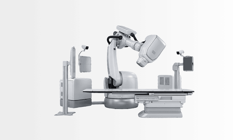
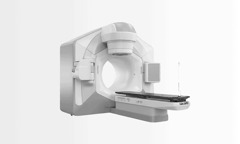
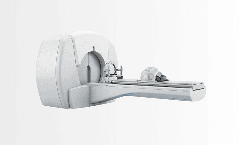
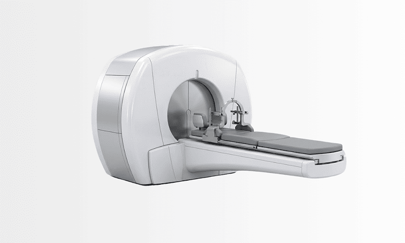
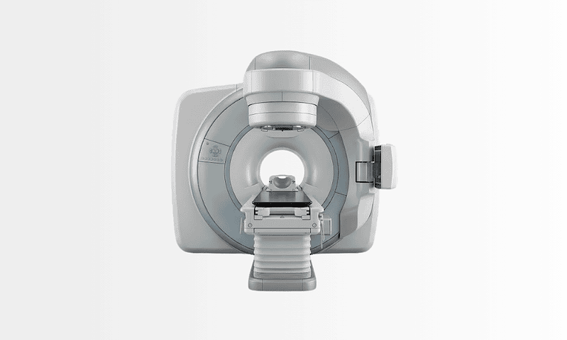
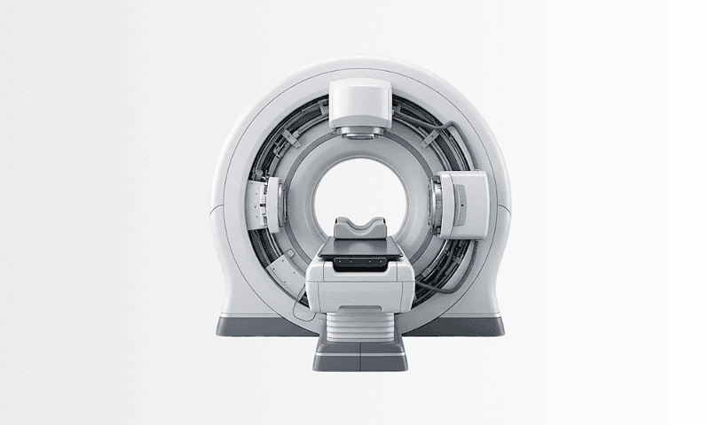
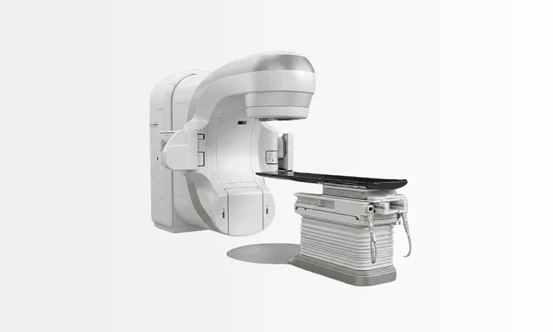
Available at these Hospitals










What patients say about this unit
★★★★★From 2,400+ verified patient reviews“From the first consultation to discharge, my targeted radiation therapy went smoothly. Asst. Prof. Dr. Güral was thorough, professional and genuinely caring. I only wish I had come here sooner.”
“My experience with oncology consultation here restored my faith in medical care. Assoc. Prof. Şahin explained every step clearly and never rushed me. I felt looked after at every turn.”
“My radiation treatment exceeded every expectation I had. Assoc. Prof. Yücel answered all of my questions with patience. The nursing staff were attentive and kind throughout.”
“My radiation treatment was handled with real skill and genuine care. Dr. Can made a stressful situation feel completely manageable. I felt looked after at every turn.”
Patient stories from this unit
 Bulgaria Limb-Saving Surgery Instead of Amputation: Svetoslav’s Story
 Facing a below-knee amputation at home, Svetoslav’s leg was saved by Acibadem’s limb-salvage surgery, opening the door to an independent, prosthesis-free life.
Read the story → Netherlands Breast Cancer Second Opinion: Maria’s Story from Rotterdam
 When Maria, a 52-year-old teacher from Rotterdam, was diagnosed with breast cancer, a rapid second opinion at Acibadem Oncology Centers changed her treatment path…
Read the story →Comparison guides for this specialty
Speak with our medical team
Share your case and our international patient team will guide you to the right specialist, free of charge.