

Dermatology
Medical and cosmetic dermatology, mole checks and skin cancer surgery, acne, eczema and psoriasis care, laser treatment, with the hospital's own dermatopathology laboratory behind every biopsy.
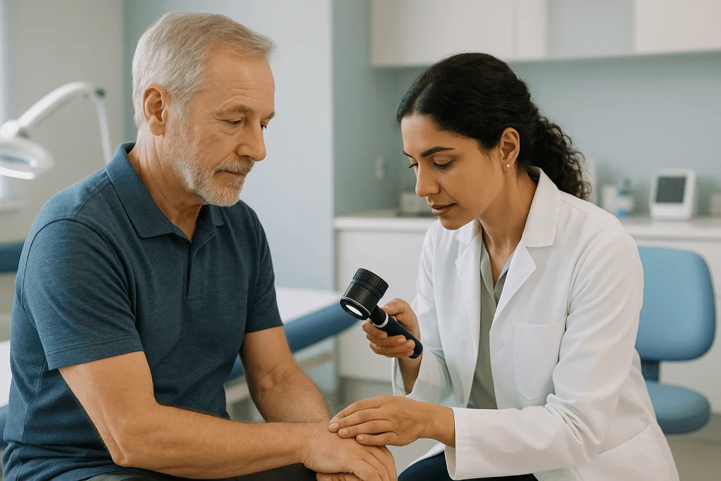
Medical, surgical and cosmetic dermatology
The same department handles the rash that will not settle, the mole that changed, and the treatment you chose for yourself, because separating them is exactly how a melanoma gets lasered off as a blemish.
Medical dermatology
Long-term skin, hair and nail disease, diagnosed properly and managed over time rather than suppressed.
Skin cancer and surgery
Mole checks, dermoscopy and excision, with our own dermatopathology laboratory reading the tissue.
Cosmetic dermatology
Laser, injectable and resurfacing treatment, offered where it fits and declined where it does not.
The laboratory is the whole argument
Most dermatology looks identical from the outside: a room, a light, a doctor. What separates a hospital department is what happens after a lesion is removed, whether the tissue reaches a dermatopathologist who reads skin all day, and how quickly the answer comes back.
It is also what happens when the diagnosis is not skin-only: a rash that turns out to be a drug reaction, a lesion that needs oncology, a child who needs a paediatrician in the same building.
What we will not do
- Laser, burn or freeze a pigmented lesion that has not been examined with a dermatoscope.
- Promise a cure for eczema, psoriasis, rosacea, vitiligo or alopecia areata.
- Quote a course of laser sessions before seeing the skin and the hair.
- Treat a changing mole as a cosmetic request.
- Start isotretinoin without the monitoring and contraception discussion that goes with it.
Who would actually see you
Every dermatologist has a profile you can read before any appointment.
How a skin problem is worked up when you are travelling
Send photographs and history
Clear images of the lesion or rash, what changed and when, and any treatment already tried.
Dermatologist review
A specialist tells you what can be assessed remotely and what genuinely needs an examination.
Examination and dermoscopy
Full-skin examination, dermoscopy of anything suspicious, and biopsy the same visit where needed.
Pathology and the plan
Tissue goes to our own dermatopathology laboratory; the report decides what happens next.
Treatment and follow-up
Courses that need several sessions are scheduled around your travel, with review after you fly home.
Six things a skin clinic brochure leaves out
A changing mole is not a cosmetic question
Any mole that changes shape, colour or size, bleeds, itches or looks different from your others needs assessment, not a laser.
Nothing pigmented is burned or frozen
A lesion destroyed by cautery or cryotherapy cannot be examined afterwards. Anything uncertain is excised and sent to pathology.
Laser is hair reduction, not removal
A completed course leaves hair sparser and finer. Hormonal change can reactivate follicles, and white or very fine hair does not respond.
Chronic disease is controlled, not cured
Eczema, psoriasis, rosacea and vitiligo are managed over time. Anyone promising a cure is selling something.
Isotretinoin needs monitoring
It is effective and it is not casual: pregnancy is an absolute contraindication, and bloods and mood are followed throughout.
Some rashes are emergencies
A widespread rash with fever and mucosal involvement, or a spreading red area with fever, needs assessment the same day.
Jump to what you came for
Quick answer
Dermatology is the medical specialty that diagnoses and treats conditions affecting the skin, hair, nails, and related mucous tissues, from common rashes and acne to chronic inflammatory and autoimmune disorders. At Acibadem in Turkey, dermatology care includes specialist evaluation, diagnostic testing when needed, and personalized treatment plans such as medical therapy, procedural care, and follow-up according to the patient’s condition.
Dermatology covers two things that look unrelated and are not: the medical care of skin, hair and nail disease, eczema, psoriasis, acne, infections, skin cancer, and the treatments people choose for how their skin looks. At Acıbadem International both sit inside a hospital department, which matters most in the place patients think about least: what happens to the tissue after a lesion is removed, and who is in the building when a rash turns out to be a drug reaction rather than an allergy.
This guide explains how skin disease is actually diagnosed, what each treatment does and does not do, and what a course of treatment looks like when you are travelling for it. Where the honest answer is “it depends on what the examination shows”, we say exactly that.
What a hospital dermatology department is, and how it differs from a skin clinic
Dermatology has two arms that are often sold as one. Medical dermatology diagnoses and treats disease of the skin, hair and nails, acne, eczema, psoriasis, urticaria, infections, hair loss, and skin cancer. Cosmetic dermatology treats the appearance of skin that is not diseased, lines, pigmentation, unwanted hair, scars, texture. Both are legitimate. The problem is that on the open market they are usually delivered by the same device, in the same room, by people with very different training, and the patient has no way to tell which one they are getting.
In a hospital department the two arms sit on top of the same medical infrastructure, and that is the practical difference. When a lesion is removed here, it goes to a dermatopathology laboratory in the same institution and comes back as a written report with a diagnosis, a measured depth where that applies, and a comment on whether the margins are clear. When a mole is photographed under dermoscopy, the image is stored in the hospital record, so the comparison at your next visit is against your own skin rather than someone’s memory. When a rash turns out to be the first sign of a systemic illness, or a biopsy turns out to be melanoma, the next specialist is down the corridor and working from the same file.
What that buys you, concretely:
- An on-site pathology laboratoryevery excised lesion is examined, not discarded. A skin specialist who cannot tell you where your tissue was sent is not offering you the same service.
- Operating theatres and anaesthesiafor lesions too large, too deep or too awkwardly placed for a treatment room, and for patients whose other conditions make a minor procedure not minor.
- Other specialties in the same buildingoncology, plastic and reconstructive surgery, rheumatology, endocrinology, paediatrics, immunology. Skin disease crosses into all of them.
- Medication safety infrastructurelaboratory monitoring for isotretinoin, screening before biologics and JAK inhibitors, and infusion facilities where a treatment needs them.
- A permanent recorddermoscopy images, pathology reports and treatment history that a second opinion anywhere in the world can read.
A well-run aesthetic clinic can do good cosmetic work. What it cannot easily do is tell you that the thing you came in to have lasered should have been biopsied instead. A full skin examination also sits naturally alongside a wider health assessment such as a check-up programme.
The skin, hair and nail problems that belong with a specialist, not a pharmacy shelf
Most skin complaints settle on their own or with something bought over the counter, and it would be dishonest to pretend otherwise. The useful question is not “is this serious?” but “how long should I reasonably wait before this needs a dermatologist?” Skin is the one organ you can watch directly, which makes waiting sensible, and makes waiting too long unusually easy.
The problems that belong with a dermatologist rather than with continued self-treatment:
- A rash has lasted more than six weeks, keeps returning to the same place, or is spreading despite treatment. Six weeks is also the line at which urticaria stops being acute and becomes chronic, which changes the investigation entirely.
- Acne has not improved after around three months of consistent pharmacy treatment, is deep, painful and nodular, or is already leaving marks. Scarring is far easier to prevent than to treat: this is the single most common reason people arrive too late.
- A mole or spot has changed in size, shape, colour or border, has started to itch, bleed or crust, or simply looks different from all your others. Change is the signal; a lesion that has looked the same for twenty years rarely is.
- Any lesion has failed to heal within four weeks, particularly on the nose, ear, lip, eyelid or scalp.
- Itching is severe, generalised or waking you at night without an obvious rash: this can reflect thyroid, liver, kidney or blood disease and deserves investigation rather than antihistamines indefinitely.
- Hair is shedding heavily for more than three months, thinning in a defined pattern, coming out in round patches, or the scalp is painful, scaly or scarred. Scarring hair loss is not reversible once the follicle is destroyed, so it is assessed early rather than watched.
- A nail is changingthickening, lifting, splitting, or developing a dark line running from the cuticle. A new pigmented band in a single nail is checked, not creamed.
- Anything on the skin is affecting sleep, work or mood. That is a medical reason, not a vanity one.
What does not need specialist assessment straight away: an isolated spot, mild dandruff responding to an antifungal shampoo, a small patch of dry skin in winter that improves with moisturiser, an insect bite that is settling day by day. These usually settle over a few weeks. Where they behave as expected, they were what they looked like. Where they do not, that itself is the information.
Skin problems that are genuine emergencies
Almost nothing on the skin is an emergency. A small number of things are, and they are the ones most often mistaken for a rash that can wait until Monday. The distinction that matters is not how bad it looks but whether the skin sign belongs to an illness happening in the rest of the body.
The skin presentations that are treated as medical emergencies:
- Swelling of the lips, tongue, mouth or throat, a changed voice, or difficulty swallowing or breathing. This is angioedema and it can close the airway.
- A new medication followed by painful skin, blisters, peeling, or sores in the mouth, eyes or genitals. Mucosal involvement after a new drug is the signature of Stevens–Johnson syndrome and toxic epidermal necrolysis: the most dangerous entry on this list, and the one most regularly mistaken for an allergy that can wait.
- A rash that is widespread and comes with fever, feeling profoundly unwell, or swollen glands. Severe drug reactions and systemic infections present this way.
- Redness spreading with heat, swelling, pain and fevercellulitis or erysipelas. Pain out of all proportion to what the skin looks like, or an area advancing over hours, raises the possibility of a deep, rapidly spreading infection. The same signs carry more weight in diabetes, lymphoedema, or a suppressed immune system.
- A child with fever and a rash that does not fade when pressed under a glass or a finger.
Shingles involving the eye area or the tip of the nose, shingles that is widespread, and shingles in anyone whose immune system is suppressed are treated urgently: antiviral treatment is far more useful when it is started within the first days, and eye involvement risks sight.
A mole that is changing, itching or bleeding, a lesion that is growing or has failed to heal in four weeks, and a new dark band under a nail belong in a dermatology clinic rather than an emergency department. Finding them earlier rather than later is where the outcome is decided.
How skin disease is actually diagnosed: dermoscopy, patch tests, biopsy and the lab behind them
Dermatology looks like a visual specialty and is not one. A great deal of skin disease looks alike, psoriasis and eczema on a leg, a benign keratosis and an early skin cancer on a temple, a fungal rash and a drug reaction on a trunk, and the treatments diverge sharply. A steroid cream applied to a fungal infection makes it worse and disguises it. An antifungal applied to eczema does nothing for months. Getting the diagnosis right is not a formality before treatment; it is most of the work.
The clinical examination. A proper dermatology consultation involves undressing to underwear and being examined under good light, including the scalp, behind the ears, between the toes, the nails and, where relevant, the mouth and genital skin. Patients frequently come in about one lesion and leave with a different one having been found. A list of every medication and supplement taken, including anything started in the past three months, and photographs of the rash at its worst where it comes and goes, are part of that assessment.
Dermoscopy. A dermatoscope is a magnifying lens with polarised light that shows pigment, vessel patterns and structures beneath the surface of the skin. In trained hands it substantially improves the accuracy of deciding which lesions need removing and, just as importantly, which do not, sparing a great many unnecessary excisions. Digital dermoscopy stores the images, so a borderline lesion can be re-photographed at three, six or twelve months and compared with what it looked like before. Comparison over time is a diagnostic tool in its own right, and it only exists if someone kept the first image.
Skin biopsy. Taken under local anaesthetic in a treatment room, usually in under twenty minutes. A shave biopsy removes a raised lesion at surface level. A punch biopsy takes a small cylinder of full-thickness skin, typically closed with one or two stitches. An excisional biopsy removes the whole lesion with a margin and is what a pigmented lesion suspicious for melanoma requires, because depth cannot be measured on a fragment. You will feel the anaesthetic injection sting; after that you should feel pressure but not pain.
The laboratory. The specimen goes to dermatopathology, where it is processed, stained and read by a pathologist. The report names the diagnosis, and for a cancer it states the type, the thickness or depth, features such as ulceration, and whether the margins are clear. Additional stains and direct immunofluorescence, used for blistering and autoimmune skin disease, are done on the same tissue. Results typically take several working days, longer if extra stains are needed, and difficult cases are reviewed by more than one pathologist rather than rushed. Where a report changes the plan, it is discussed with the relevant team; a confirmed melanoma, for example, moves immediately into a shared pathway with medical oncology.
Other tests. Patch testing applies a panel of common allergens to the back for 48 hours, with readings at 48 and 96 hours, to identify contact allergy: the only reliable way to find it. Skin scrapings and nail clippings for microscopy and fungal culture confirm infection before anyone commits you to months of oral antifungal treatment. Trichoscopy examines the scalp; blood tests screen for the internal causes of itching, hair loss and urticaria. Photographs, with your consent, document what changes.
Acne: from the first spot to isotretinoin, and when scarring becomes the real problem
Acne is a disease of the hair follicle and its oil gland, driven by blocked follicles, oil production, bacteria and inflammation, usually with a hormonal engine behind it. It is not caused by poor hygiene, and scrubbing harder reliably makes it worse. It is also not trivial: it occurs on the face, at the age when people are forming a sense of themselves, and it can leave permanent marks. Treating it properly is medicine, not cosmetics.
Severity guides the treatment, and so does scarring. Comedonal acne, blackheads and whiteheads without much inflammation, is usually managed with topical treatment. Inflammatory acne with red papules and pustules often needs a combination of topical agents, sometimes with a course of oral antibiotic. Nodular and cystic acne, with deep, painful lumps, is a different problem: it scars, and it is treated as though scarring is the outcome to be prevented rather than a possibility to be observed. So is any acne that is already leaving marks, however mild it looks. That is the point at which “let’s see how it goes” stops being reasonable advice.
The usual sequence. Topical retinoids, benzoyl peroxide, topical antibiotics used in combination rather than alone, and azelaic acid form the base. Oral antibiotics are added for inflammatory acne for a limited period, months, not years, because prolonged use drives resistance and does not address the cause. For women whose acne flares along the jaw and chin, worsens before periods, or arrives in adulthood, hormonal treatment such as the combined oral contraceptive or an anti-androgen is often more effective than antibiotics; where acne comes with irregular periods, unwanted facial hair or scalp thinning, polycystic ovary syndrome and other endocrine causes are investigated rather than assumed. Doses and choices belong to the prescribing doctor and depend on your history, other medicines and pregnancy plans.
Isotretinoin. For severe, scarring or genuinely treatment-resistant acne, oral isotretinoin is the most effective option available, and for many patients a full course produces lasting remission. It also demands respect. It is absolutely contraindicated in pregnancyit causes serious birth defects, so effective contraception and a pregnancy prevention programme with regular testing are mandatory before, during, and for a defined period after treatment for anyone who could become pregnant. It requires blood tests for liver function and lipids during the course. It dries skin, lips and eyes, and can make skin fragile: waxing, laser treatments and other resurfacing are postponed until an interval after the course has finished. Mood changes are discussed openly at the start and monitored throughout, and they are reported to the prescriber rather than left until the next appointment. Acne often flares in the first weeks before it improves. None of this makes isotretinoin unsafe; it makes it a drug that belongs under supervision with a laboratory attached.
What is not acne. Itchy, uniform small bumps on the chest, back and shoulders that respond poorly to antibiotics are often fungal folliculitis, which antibiotics can aggravate. Persistent central facial redness with flushing and papules is more likely rosacea. Painful recurrent lumps and tunnels in the armpits, groin or under the breasts are hidradenitis suppurativa: a distinct inflammatory disease with its own treatments, frequently mislabelled as boils for years. Sudden severe acne in an adult, particularly with other hormonal signs, prompts investigation rather than a stronger cream.
Scarring. Once acne has settled, what remains is usually a mixture of red or brown marks, which fade over months, and true scars, which do not. Post-inflammatory pigmentation is common in darker skin and improves with time, sun protection and pigment-directed treatment. Genuine atrophic scars, ice pick, boxcar and rolling, need physical treatment, and the honest position is that scars can be improved, sometimes considerably, but not erased. How each type is treated, and what a realistic result looks like, is set out under skin quality and resurfacing below.
Immune-driven skin disease: eczema, psoriasis and chronic urticaria
Eczema, psoriasis and chronic urticaria are not infections, they are not caused by poor hygiene, and you cannot catch them or pass them on. They are conditions in which the immune system reacts against the skin’s own tissue, which is why they flare, settle and return. That shapes the honest goal of treatment: these diseases are controlled and pushed into long, comfortable remissions. They are not cured by a single course of anything, and a clinic promising otherwise is describing something other than the disease you have.
Eczema is an umbrella term rather than one diagnosis, and naming the type changes the treatment:
- Atopic eczemaa damaged skin barrier plus immune overactivity, usually with a personal or family history of asthma or hay fever. It favours the elbow and knee creases, neck and hands, and the itch–scratch cycle does much of the damage.
- Contact dermatitisirritant (soap, water, solvents, occupational exposure) or allergic (nickel, fragrance, hair dye, preservatives). Patch testing separates them and names the actual culprit, as described under diagnosis.
- Seborrhoeic dermatitisscalp, eyebrows, sides of the nose and chest, linked to the Malassezia yeast that lives on everyone’s skin; see scalp and nail conditions.
- Dyshidrotic and discoid eczemadeep itchy blisters on palms and soles, and coin-shaped plaques regularly mistaken for ringworm and treated, unsuccessfully, with antifungal cream.
Psoriasis is a systemic immune condition that happens to show on the skin: plaques on elbows, knees, lower back and scalp, guttate psoriasis after a throat infection, inverse psoriasis in the body folds, and nail pitting, thickening and separation from the nail bed. Joints matter here as much as skin. Morning stiffness lasting more than half an hour, a swollen finger or toe, or persistent heel pain can indicate psoriatic arthritis, and joint damage that has already happened cannot be reversed, which is why that referral is made early rather than eventually. Psoriasis also travels with higher cardiovascular and metabolic risk, so blood pressure, lipids and blood sugar belong in the assessment.
Psoriasis or eczema? In eczema, itch dominates, the edges of the rash are poorly defined, the skin weeps or crusts, and the flexures are typically involved. In psoriasis, plaques are sharply demarcated with thick silvery scale, prefer the extensor surfaces, scalp, navel and natal cleft, may itch less but burn or crack, and the nails often give the diagnosis away. The two overlap in real patients, and where the picture is mixed a biopsy settles it rather than a longer trial of the wrong cream.
Chronic urticaria means hives that keep appearing for more than six weeks. Most cases are chronic spontaneous urticaria, in which no external trigger is ever found, extensive food-allergy testing usually adds cost rather than answers. Antihistamines are the starting point, at a dose adjusted by a doctor rather than by you, and further options including a biologic exist when they are not enough. One warning belongs here: swelling of the lips, tongue or throat, a change in the voice, or any difficulty swallowing or breathing is angioedema affecting the airway, which is a medical emergency rather than a dermatological one.
Treatment climbs a ladder, emollients and topical steroids matched in strength to the body site, calcineurin inhibitors for face and folds, narrowband UVB phototherapy, then systemic drugs, biologics and JAK inhibitors for severe or unresponsive disease. Undertreating a flare with too weak a steroid for too short a time is at least as common as overusing one. Before any systemic drug, screening for tuberculosis, hepatitis and other infections plus baseline blood tests is mandatory, with monitoring afterwards, work we do alongside internal medicine. Biologics are continuing therapies, not a holiday course, so supply and follow-up at home are arranged before treatment starts and the patient’s own doctor is written to. Stopping them usually means the disease returns.
Redness and pigment: rosacea, melasma, sun spots and vitiligo
These conditions are usually filed under “cosmetic”, which is exactly why they are so often treated badly. Facial redness and pigment patches are medical diagnoses with medical assessments behind them, and the wrong product makes several of them permanently worse. The clearest example is a steroid cream on a red face: it looks like a good decision for about a week, then produces rebound redness, pustules and, with continued use, steroid-induced rosacea or perioral dermatitis.
Rosacea is not adult acne and is not a hygiene problem. It appears in overlapping patterns, persistent central facial redness with visible thread veins; a papulopustular pattern with inflamed bumps and pustules but none of the blackheads that define acne; a phymatous pattern in which the skin of the nose thickens over years, most often in men; and ocular rosacea, with gritty, dry, red eyes and inflamed lid margins, which is missed constantly because patients do not connect their eyes to their skin. Persistent eye symptoms are assessed by an eye specialist alongside skin treatment. Management combines trigger awareness, sun, heat, alcohol, spicy food and stress affect people differently, so a short diary beats a generic avoidance list, with prescribed topical and oral treatment. Fixed dilated vessels and background redness do not respond to creams and are treated with vascular laser, described under pigment and vascular lasers.
Melasma is symmetrical brown or grey-brown patching across the cheeks, forehead and upper lip, driven by hormones (pregnancy, combined contraception), ultraviolet light, visible light and heat. The honest framing is that melasma is managed, not deleted; it recurs whenever photoprotection lapses, which is why daily broad-spectrum sunscreen, tinted, containing iron oxides, because visible light also drives it, matters more than any device. Prescribed topical combinations work but are supervised, since prolonged unsupervised use of some of them causes its own pigment problems. Peels and certain lasers help selected patients and worsen melasma in others, particularly in darker skin. Anyone offering to clear melasma permanently in one laser session is not describing melasma.
Two other pigment problems come up constantly. Post-inflammatory hyperpigmentation is the brown mark left by acne, eczema or a procedure: not a scar, fading over months rather than weeks, and more persistent in darker skin. Solar lentiginesthe flat brown sun spots on face, chest and backs of the hands, are cumulative sun damage. One caution outranks every cosmetic consideration here: a pigmented patch that is enlarging, developing uneven colour, itching or bleeding is never assumed to be a harmless sun spot. It is examined with dermoscopy and, where there is any doubt, removed and sent to pathology, see melanoma.
Vitiligo is an autoimmune condition in which pigment cells are lost, leaving sharply defined white patches, often symmetrically and often first on the hands and around the eyes and mouth. Because it keeps company with other autoimmune conditions, thyroid function is usually checked. Options include topical steroids and calcineurin inhibitors, narrowband UVB, targeted excimer light for limited patches, and newer topical treatments. Repigmentation is slow, measured in months of consistent treatment, and uneven: the face and neck respond best, hands and feet least, results vary considerably between individuals, and relapse after a good response is possible. There is no cure, and being told otherwise is a reason to be sceptical. Depigmented skin has no protection against ultraviolet light, so sun protection is part of treatment rather than an afterthought, cosmetic camouflage is a legitimate option, and the psychological weight of a visible pigment condition is taken seriously here rather than dismissed as vanity.
Skin infections: fungal, viral and bacterial
Skin infection is where self-treatment goes wrong most often, and usually in the same way: a rash is treated with a steroid cream, the redness improves briefly, and the fungus underneath spreads under cover, tinea incognito, a rash that no longer looks like anything recognisable by the time it reaches us. The first job is to name the organism rather than guess it. Skin scrapings for microscopy and culture, bacterial swabs, viral testing where it changes management and occasionally a biopsy are quick, and they prevent months of the wrong treatment.
Fungal infections are common and mostly straightforward once identified:
- Tinea (ringworm) of the body, groin and feetusually clears with a topical antifungal used for the full recommended duration, not until it stops itching; extensive disease needs oral treatment.
- Tinea capitis in childrenscalp ringworm cannot be cleared by shampoo or cream alone. It requires oral treatment, and household members are often checked, because reinfection is otherwise routine; children’s skin is covered with our paediatrics colleagues.
- Pityriasis (tinea) versicolorpale or darker patches on the trunk and shoulders, most obvious after sun exposure. It recurs in warm weather, and the colour takes months to even out after the yeast has gone, which is not treatment failure.
- Candida in skin foldsrecurrent or extensive cases prompt a check for diabetes and other predisposing factors.
- Nail fungusnever started on appearance alone. Months of oral antifungal treatment, with liver monitoring and drug interactions to consider, are justified only after microscopy or culture confirms it; see scalp and nail conditions.
Viral infections ask for patience more than firepower. Warts are caused by human papillomavirus and often need repeated cryotherapy, topical treatment or curettage over months, plantar warts being the most stubborn; the threshold for treatment is lower if your immune system is suppressed. Molluscum contagiosum in children resolves on its own over months, and aggressive treatment of every lesion tends to leave marks that outlast the virus, so watchful waiting is frequently the better medicine. Herpes simplex responds best to antiviral treatment started at the first tingle rather than once blisters are established. Shingles has clear red lines: a rash around the eye or on the tip of the nose is urgent because the eye can be involved and sight is at risk, and shingles that is widespread or occurring in someone immunosuppressed is urgent as well. Antivirals do most of their work when started within roughly 72 hours of the rash appearing, and nerve pain persisting long after the rash has healed is a recognised complication managed with our pain management team rather than endured.
Bacterial infections range from trivial to genuinely dangerous. Impetigo, golden-crusted patches, common in children, is contagious and needs treatment plus a short period away from school or work. Folliculitis and boils are usually simple, but recurrent boils are swabbed rather than treated blindly again and again. Hidradenitis suppurativa, with recurrent painful nodules and tunnels in the armpits, groin and under the breasts, is not poor hygiene and is not a run of ordinary boils; it is an immune-driven condition, routinely diagnosed years late, with proper medical and surgical treatment pathways. Cellulitis is the one to know: spreading redness with warmth, swelling, tenderness and fever is a bacterial infection of the skin that needs medical treatment, and it carries more weight still with diabetes, lymphoedema or a suppressed immune system. Pain far out of proportion to what the skin looks like, an area advancing over hours, or feeling severely unwell are the features that mark out the dangerous end of this spectrum.
Restraint counts too. Not every red, warm area is infected, stasis dermatitis, contact dermatitis and inflamed cysts are misdiagnosed as cellulitis routinely, and redness affecting both legs symmetrically is rarely cellulitis at all.
Skin cancer: the three types that behave completely differently
“Skin cancer” is three different diseases with three different personalities, plus a precursor lesion that can be treated before it becomes anything. Basal cell carcinoma grows locally over years and almost never spreads elsewhere. Squamous cell carcinoma grows faster and can spread to lymph nodes. Melanoma, dealt with in the next section, is far less common and behaves differently again. What they share is this: an early lesion is dealt with completely by removing a small piece of skin, while the same cancer left for years destroys the nose, eyelid or ear it grew on and demands much bigger surgery.
Basal cell carcinoma is the most common cancer in humans. It typically appears on the face, nose, cheeks, eyelids, ears and scalp as a pearly or translucent bump with fine visible blood vessels across it and a rolled edge, sometimes with a central crust or dip; a flatter, scar-like form exists and is easy to overlook. The classic history is a sore that bleeds slightly, scabs over, seems to heal and then breaks down again in the same spot over months. It rarely spreads to other organs but invades locally into cartilage and bone if ignored. Treatment is surgical removal with a margin; for high-risk facial sites, Mohs surgery removes it with immediate margin checking, described under skin surgery. Selected superficial lesions are treated with curettage, cryotherapy or topical field treatment, and radiotherapy is an option where surgery is not suitable.
Squamous cell carcinoma looks different: a scaly, crusted, often tender nodule growing over weeks to months, frequently on the lip, ear, scalp, forearms or backs of the hands. Because it can spread to lymph nodes, it is treated promptly rather than watched. The risk rises sharply and permanently after organ transplantation and during long-term immunosuppression, which is why scheduled skin surveillance is used in that group rather than waiting until something is noticed.
Actinic keratoses are the precursor stage: rough, dry, sandpapery patches on the scalp, face, ears and hands, often easier to feel than to see. Some progress to squamous cell carcinoma and there is no way to tell in advance which, so they are treated, cryotherapy for individual lesions, topical field treatment or photodynamic therapy when a whole sun-damaged area is involved. Treating the field rather than one spot is the point.
Risk concentrates in fair skin that burns easily, childhood sunburn and sunbed use, outdoor work, long-term immunosuppression, previously irradiated skin, chronic wounds and old scars, and above all in anyone who has already had one skin cancer. Darker skin is not exempt: skin cancer in brown and black skin is less common but diagnosed later, and more often arises in less sun-exposed sites: the soles, palms, under the nails and inside the mouth.
One pattern deserves separate mention because it is repeatedly treated as dermatitis. A scaly, itchy, weeping change of the nipple and areola on one side that does not settle with eczema treatment may be Paget’s disease of the nipple, which is associated with underlying breast cancer. Persistent one-sided nipple changes are assessed with our breast health team rather than given another cream.
The practical rule: any skin lesion that has not healed within four weeks, or that keeps bleeding, is examined rather than watched. No photograph and no examination alone establishes the diagnosis; a biopsy and a pathology report do. Where disease is advanced, recurrent or inoperable, decisions are made in a tumour board with medical oncology, dermatology, pathology and surgery in the same room.
Melanoma: the ABCDE rule, the subtypes that hide, and why staging decides everything
Melanoma is much less common than the other skin cancers and accounts for most deaths from skin cancer, because it can spread to lymph nodes and organs. Almost everything about the outlook is decided by how deep it has grown into the skin at the moment it is removed, and that depth is a function of how early it was found. This is the whole argument for knowing your own skin and having changing lesions examined rather than photographed and forgotten.
The ABCDE rule is the standard starting point: Asymmetry, irregular Borders, uneven Colour with more than one shade, Diameter over about 6 mm, and: the most important letterEvolution, meaning any change in size, shape, colour, surface or sensation. Alongside it sits the “ugly duckling” sign: most of your moles resemble each other, and the one that looks unlike its neighbours deserves attention regardless of the checklist. The trap is that one dangerous subtype breaks all of those rules. Nodular melanoma is often small, symmetrical, evenly coloured and sometimes pink rather than dark, and it grows deep quickly. For that pattern the shorthand is EFG: Elevated, Firm, Growing steadily over weeks. A new lump enlarging over a few weeks deserves examination whatever its colour.
- Superficial spreading melanomathe most common form; flat or slightly raised, irregular in outline and colour, changing over months to years.
- Nodular melanomaraised, firm, fast, frequently described as having appeared and grown within weeks; it may be amelanotic, carrying little or no pigment.
- Lentigo malignaa slowly enlarging flat brown patch on chronically sun-damaged skin, usually the face of an older person, regularly dismissed as an age spot.
- Acral lentiginous melanomaon the palms, soles and under the nails. It is unrelated to sun exposure and matters most if you have brown or black skin, because it is the subtype most often found late. A new dark band running the length of a nail, especially if it is widening, irregular, in a single nail, or the pigment extends onto the surrounding skin fold, is examined properly; see nail conditions.
- Rarer forms arise in the eye and in mucous membranes, which is why unexplained pigmented spots in the mouth or genital skin are not ignored.
How fast melanoma spreads depends entirely on which of those it is. Superficial spreading melanoma may grow sideways within the top layer of skin for months or years before becoming invasive; nodular melanoma can become deep within a few months. Since neither you nor we can tell which from the surface alone, a lesion that has definitely changed is not put on indefinite observation. It is examined with dermoscopy, and where doubt remains it is removed and sent to pathology. “Let us watch it for a year” is not an acceptable answer to a mole that has already changed.
When melanoma is suspected, the lesion is removed whole with a narrow margin, an excisional biopsy, rather than shaved or partially sampled, because the pathologist has to measure how deep it goes. The report states the Breslow thickness, whether the surface is ulcerated, the mitotic rate and whether the margins are clear. Those findings drive everything after: a wider excision with a margin determined by thickness, and a discussion about sentinel lymph node biopsy when thickness and other features cross accepted thresholds. Be clear about what that node biopsy is for: it establishes the stage of the disease and informs follow-up and further treatment decisions, rather than being a treatment in its own right. Imaging is added when the stage warrants it, not routinely for thin lesions.
Beyond surgery, melanoma treatment has changed substantially: testing the tumour for BRAF and other mutations opens the door to targeted therapy, and immunotherapy with checkpoint inhibitors is used both for advanced disease and, in selected higher-risk cases, after surgery. Those decisions are made jointly with medical oncology in a tumour board rather than by any one doctor. Afterwards, follow-up is structured: scheduled full-skin examinations, because having had one melanoma raises the risk of another; instruction in examining your own skin between visits; serious sun protection; and screening for first-degree relatives where the family history warrants it. What that appointment involves in practice is set out under mole checks and skin screening.
Mole checks, mole mapping and full-body skin screening
A mole check is not a glance at the one spot that worries you. A proper skin check is a full-body examination in good light, with your clothing removed and a gown provided, covering scalp, behind the ears, the back, between the toes, the nails and, with your consent, the skin folds and genital area, because melanoma appears in places the sun never reaches and in places you cannot see yourself. Most people who book a mole check come in about a single lesion and leave with information about a different one they had never noticed.
What happens at a full body skin exam is straightforward:
- History first. Family history of melanoma, previous skin cancer, sunburns and sunbed use, fair skin that burns easily, a large number of moles, immunosuppression after transplant or for autoimmune disease. These decide how often the examination is repeated.
- Dermoscopy. Each suspicious lesion is examined with a magnifying, polarised device that shows pigment patterns and vessels invisible to the naked eye. This is where an atypical mole is separated from an ordinary one.
- Digital records. Lesions worth watching are photographed and measured, so that at your next visit the dermatologist compares images rather than memories. This comparison over three, six or twelve months is what mole mapping actually means.
- A plan per lesion. Reassure and leave, photograph and review, or remove and send to pathology. You should leave knowing which category each concerning spot falls into.
Screening reduces the chance of a late diagnosis. It does not eliminate it. Nodular and amelanotic melanomas can grow between visits and do not always follow the ABCDE pattern, which is why what happens between appointments matters more than the interval itself: a mole that changes, itches persistently, bleeds even once, or simply looks unlike all the others is assessed rather than watched until the next scheduled visit.
A dysplastic nevus, an atypical mole under the microscope, is not cancer. It is a signal that your skin makes irregular moles and that you belong in regular surveillance, sometimes with the margins of that particular lesion widened. People are often told the opposite by well-meaning relatives, which is why the pathology report is read to you rather than filed.
For anyone with fair skin, many moles or a family history, a full skin examination belongs in a general health assessment as routinely as blood pressure does.
Mole removal, cyst removal and minor skin surgery, and Mohs for skin cancer
A mole removal, a cyst removal or a lipoma excision is a small operation with a disproportionately large consequence attached to it: what the laboratory says about the tissue afterwards. That is the part a beauty setting cannot provide, and it is the reason these procedures sit inside a hospital department rather than beside a facial.
Most skin surgery is done under local anaesthetic in a treatment room, takes well under an hour, and you walk out afterwards. What separates it from the same procedure offered in a beauty setting is not the skill of the hands: it is what happens to the tissue afterwards, and who is available if the result is unexpected.
The rule we do not bend: every lesion we remove by excision goes to pathology, and nothing pigmented or uncertain is ever burned or frozen without a dermoscopic assessment first Not the suspicious ones, not the ones the patient asks about, all of them. Melanoma has been diagnosed from lesions removed because a shirt collar rubbed them, and it cannot be diagnosed from something that went into a bin. This is also why a mole should not be burned, frozen or lasered away without a diagnosis: the tissue is destroyed and the answer with it.
- Shave versus excision. A raised, clearly benign lesion can be shaved flush with the skin, healing as a flat pale mark. Anything pigmented or uncertain is excised full-thickness with a margin and closed with sutures, because the pathologist needs the base of the lesion, not just its top.
- Epidermoid and pilar cysts. A sebaceous cyst recurs when its capsule is left behind, so the aim is removal of the sac intact. A cyst that is currently red, hot and painful is usually drained and settled first, then excised later, operating through active inflammation is what produces the worst scars and the highest recurrence.
- Lipomas. Soft, mobile fatty lumps. Small ones come out through a short incision; large, deep or rapidly growing ones are imaged first, because size and growth change the differential diagnosis.
- Skin tags and seborrheic keratoses. Genuinely benign, removed by snip, cautery or cryotherapy, usually in one visit. A pigmented lesion that “looks like a seborrheic keratosis” is confirmed by dermoscopy before anything is destroyed.
For skin cancer, the operation is defined by the diagnosis. A basal cell carcinoma on the trunk may need only a modest margin. A wide local excision follows a melanoma, with the margin decided by the depth on the first pathology report. Mohs surgery, removing tissue in stages and mapping the margins under the microscope while you wait, is used where tissue must be preserved and clearance must be certain: nose, eyelid, ear, lip. It takes most of a day, not twenty minutes, and that is the point.
Every excision leaves a scar. The honest aim is the smallest, best-placed, best-settled scar, not an invisible one, and a history of bad scarring or keloids changes that planning from the outset. Where closing the defect needs a flap or a graft, or where an old scar needs revising, the operation is planned together with plastic surgery from the outset rather than as a rescue.
Laser hair removal: what the machine can and cannot do
Laser hair removal works by heating pigment in the hair follicle, which means it only affects hairs that are dark and currently in their growing phase. At any moment, only a fraction of your follicles are in that phase, which is why this is a course of treatments spaced weeks apart, not an appointment. Anyone who tells you the exact number of sessions you will need before seeing your skin and hair is guessing; it depends on the area, hair colour and thickness, your hormones and how you respond.
The correct term is permanent hair reduction, and it is used deliberately. A well-run course leaves the area with fewer, finer, lighter hairs that grow more slowly, and most people need occasional maintenance afterwards. Grey, white, red and very fine blond hairs contain too little pigment for the laser to target and will not respond, whatever the device. If someone promises permanent removal of all hair, they are describing something the technology does not do.
- The wavelength is chosen for your skin, not for marketing. Alexandrite lasers are efficient on fair skin with dark hair. Diode sits in the middle and covers large areas quickly. Nd:YAG is the safer choice for brown and black skin (Fitzpatrick IV–VI) because it passes deeper and interacts less with pigment in the epidermis. Matching device to skin type is the single decision that prevents burns and pigment change.
- IPL is not a laser. Intense pulsed light is a broad-spectrum lamp; it can reduce hair, less selectively and usually less durably. It is a legitimate tool, but it should be named accurately.
- Electrolysis is the alternative for pale hair. Slower and hair-by-hair, but it does not depend on pigment: the reason it still exists.
- Preparation matters. The area is shaved rather than waxed, plucked or threaded for several weeks beforehand, and skin that is tanned or freshly self-tanned is not treated. Tanned skin raises the risk of burns and blistering, so treatment is postponed rather than pushed through.
Medication matters here. Isotretinoin, some antibiotics and other photosensitising drugs change how skin reacts to laser, and treatment is timed around them. Active infection, certain skin conditions in the treatment area and pregnancy also change the plan.
It is not painless. Most people describe a hot elastic snap, worse on the upper lip, bikini area and shins; cooling and settings make it tolerable, and areas such as the underarm take minutes. Expect redness and follicular swelling for a day or two. The real risks, blistering, burns, and temporary or, rarely, lasting lightening or darkening of the skin, come almost entirely from the wrong wavelength or energy on the wrong skin, which is why the assessment matters more than the brand name on the machine.
One medical caveat: facial or body hair that has increased noticeably alongside irregular periods, acne or scalp thinning is a symptom rather than a cosmetic problem. Hormonal causes are investigated before a laser course begins, because treating the hair without treating the cause means it keeps coming back.
Botox, fillers and skin boosters: what they do and where they stop
Injectables are medical treatments with medical risks, and they are the area where the gap between a hospital dermatology clinic and a hotel-room appointment is widest. They work on specific problems, they wear off, and they do not do the job of surgery.
Botulinum toxin relaxes the muscles that fold the skin, forehead, frown, crow’s feet, and in trained hands the jaw, neck bands and excessive sweating. It softens lines caused by movement. It does not fill volume, tighten loose skin or lift a heavy brow beyond a few millimetres. The effect appears over several days and fades gradually over a few months, sooner in strong muscles and with very small doses. Bruising and temporary asymmetry happen; a droopy eyelid or brow is uncommon, settles as the effect wears off, and is far more likely when the injector does not know the anatomy underneath.
Hyaluronic acid fillers replace volume and support: cheek, chin and jawline structure, nasolabial folds, lips, and, with great caution, tear troughs, where fluid retention and a bluish shadow are common complications and where many people are better served by doing nothing. Their advantage over permanent materials is that they can be dissolved with hyaluronidase if the result is wrong or the product migrates.
The complication you must know about before you consent: filler injected into or compressing a blood vessel can block the blood supply to skin, and, in the areas around the nose, glabella and eyes, to the eye itself. That means tissue loss and, rarely, permanent visual loss. It is treatable only if it is recognised immediately, which is why the warning signs are part of the consent conversation before any injection:
- Severe pain out of proportion to the injection, immediately or in the hours afterwards.
- Blanching, white or dusky mottled skin anywhere near the treated area.
- Any change in vision, or pain in or behind the eyethis is an emergency.
Skin boosters and polynucleotides are different again: injected hydration and stimulation spread across an area, aimed at skin quality rather than shape, over a short series. Thread lifts reposition tissue modestly and temporarily, with threads that dissolve. All of these have a ceiling. When the underlying issue is skin laxity and descent rather than volume or lines, more filler makes a face heavier, not younger: the honest advice at that point is a surgical assessment with plastic surgery, and we will say so rather than sell another syringe.
Peels, microneedling and resurfacing lasers, including acne scars
Everything in this group works the same way: controlled, measured injury that makes the skin repair itself with better collagen and more even pigment. The differences are depth, downtime and risk, and the honest framing is that they improve skin, they do not reset it. Recovery is real, and any treatment sold as transformative with no downtime is one or the other, not both.
- Chemical peels. Superficial peels freshen texture and mild pigmentation over a series with a few days of flaking. Medium-depth peels, such as TCA, do more for pigment and fine lines and take a week or more of visible peeling. Deep peels are powerful, unforgiving and rarely the right first answer.
- Microneedling. Fine needles create controlled channels that stimulate collagen; adding radiofrequency delivers heat deeper for laxity. Modest, cumulative, low downtime, and useful in darker skin where aggressive lasers carry more pigment risk.
- Resurfacing lasers. Non-ablative fractional lasers give gradual improvement across several sessions with days of redness. Ablative fractional CO2 does substantially more in one session and costs you a week or two of genuine healing, with strict aftercare and sun avoidance.
- Skin-quality facials. Device-assisted cleansing and hydration treatments make skin look better for an event. They are low-risk, though they are not performed over active infection or active acne; they are not scar or wrinkle treatments, and we will not describe them as such.
Acne scars deserve their own paragraph, because the type of scar decides the method and no single device treats them all. Rolling scars, tethered from below, respond to subcision, releasing the fibrous band with a needle, often combined with collagen stimulation. Boxcar scars, with defined edges, respond to fractional resurfacing. Ice-pick scars, narrow and deep, respond best to focal TCA application into the scar itself rather than to any surface laser. Most people have a mixture, so a realistic plan combines methods across several sessions over months. Post-inflammatory redness and brown marks are not scars at all and usually fade with time, sun protection and pigment-directed treatment.
Two conditions before any of this. First, active acne is treated before scars are, because resurfacing inflamed skin creates new scarring. Second, in brown and black skin every one of these treatments carries a real risk of post-inflammatory hyperpigmentation, so settings are conservative, priming and sun protection are non-negotiable, and a test area is sensible. Acne scars can be significantly improved. They cannot be erased, and anyone claiming otherwise is describing a photograph, not a result.
Pigment, vascular and tattoo lasers
Removing colour from skin is a different job from removing hair, and it uses different machines. Pigment and tattoo lasers work by releasing their energy in extremely short pulses, nanoseconds for Q-switched devices, trillionths of a second for picosecond devices, so that the pigment particle absorbs the energy and shatters before the surrounding skin has time to heat up. Vascular lasers do the opposite: they target the red of haemoglobin inside a blood vessel and heat it long enough to seal it. Neither of these lasers should ever be pointed at a pigmented lesion that has not first been examined with a dermatoscope. A laser can clear the visible surface of a melanoma while leaving the disease beneath it, and destroy the evidence that would have made the diagnosis. In this department, any brown mark being considered for laser is assessed as a lesion first and a cosmetic problem second.
- Sun spots, lentigines and freckles. Discrete brown marks caused by ultraviolet damage respond predictably to pigment lasers and to intense pulsed light, usually in a small number of sessions. What laser cannot do is stop new ones forming: without daily sun protection, they return, and freckles in fair skin reappear every summer regardless of what was done to them.
- Melasma is the exception, and the honest answer is uncomfortable. Melasma is hormonally and light-driven, and laser energy is itself an inflammatory stimulus that can make it worse or trigger rebound darkening. Treatment begins with rigorous photoprotection and topical or oral medical therapy; lasers are used cautiously, at conservative settings, in selected cases only, and relapse is the rule rather than the exception. Anyone offering to clear melasma permanently in a fixed number of sessions is describing something that does not exist.
- Facial redness and visible vessels. Vascular lasers and light devices reduce the fixed redness, broken capillaries and cherry angiomas of rosacea and photodamage. Flushing, the reactive, triggered kind, responds far less well than fixed redness, and the inflammatory papules of rosacea still need medical treatment rather than light.
- Intense pulsed light is not a laser. IPL is broad-spectrum light filtered towards a target. It is useful for redness and superficial brown pigment in fair skin, and it is a poor and sometimes harmful choice in tanned skin, in darker skin types and in melasma.
Tattoo removal is a series, not a procedure. Ink is fragmented by the laser and then carried away by your own immune system over weeks, which is why sessions are spaced. Black and dark blue inks respond most reliably; red, orange and yellow are less predictable; white, flesh-toned and some cosmetic pigments can darken paradoxically on the first pass: a particular risk with eyebrow tattoo removal and permanent makeup, which is why a test patch is done first. Amateur tattoos usually clear faster than dense professional work. We do not promise a session count before seeing the tattoo, and we do not promise that nothing will remain: a faint ghost or a textural shadow is a realistic outcome for some tattoos, and you should be told that before the first pulse rather than after the sixth. Treatment is uncomfortable, most people describe it as a hot elastic band snapping against the skin, and cooling or topical anaesthetic is used. Blistering, temporary lightening or darkening of the surrounding skin, and, uncommonly, scarring are recognised risks, and the risk of pigment change is higher in darker skin.
Hair loss: finding the cause before choosing a treatment
Hair loss is one of the few complaints where the treatment sold most aggressively is the one that should be chosen last. Different causes look superficially similar and respond to completely different things, and one group of causes, the scarring alopecias, permanently destroys the follicle if it is not caught early. So the consultation begins with diagnosis, not with a product.
- History. When it started, whether you are shedding hair or thinning without shedding, what happened three months earlier (illness, surgery, childbirth, rapid weight loss, a new medication), family pattern, styling and traction, and any scalp symptoms such as itch, burning or tenderness: the last of these often points towards a scarring process.
- Examination and trichoscopy. The scalp is examined with a dermatoscope, which shows what the naked eye cannot: variation in hair shaft thickness in patterned loss, exclamation-mark hairs in alopecia areata, and, critically, whether the follicular openings are still present. When they have disappeared, the diagnosis has changed.
- Blood tests. Ferritin and a full blood count, thyroid function, vitamin D, and, where the pattern and history justify it, an androgen and polycystic ovary work-up in women, coordinated where needed with endocrinology. Tests are chosen from the history rather than ordered as a fixed panel.
- Scalp biopsy. A small punch sample under local anaesthetic, taken when scarring alopecia is suspected or when the clinical picture is unclear. It is the only way to distinguish some of these conditions with confidence.
The common diagnoses behave very differently. Telogen effluviumdiffuse shedding a few months after a physical stress, is usually reversible once the trigger is addressed, and needs an explanation rather than a laser. Androgenetic (patterned) hair loss is progressive; medical treatment slows and partially reverses it but works only while it is continued. Alopecia areata is autoimmune and unpredictable, with spontaneous regrowth in some people and relapse in others; newer oral JAK inhibitors have changed what is possible in severe disease, and are prescription treatments requiring screening and ongoing blood monitoring. Scarring alopecias, including frontal fibrosing alopecia and lichen planopilaris, are the ones that should not wait: treatment aims to stop the inflammation and protect the hair you still have, because what has already been lost does not come back.
Medical treatments include topical minoxidil, which commonly causes a temporary increase in shedding in the first weeks before it helps, and which is often abandoned for exactly that reason, low-dose oral minoxidil in selected patients with appropriate cardiovascular assessment, and anti-androgen treatment. Finasteride is a prescription medicine with a recognised discussion around sexual side effects, and it must not be taken or handled by women who are or may become pregnant, because it can harm a developing male fetus. Platelet-rich plasma is offered here as an adjunct where it is reasonable, described honestly: the evidence is modest and mixed, the effect is not equivalent to medical therapy, and repeated sessions are needed to maintain anything gained. Surgery is a separate decision made only once the cause is established and the disease is stable, grafts placed into active scarring alopecia or untreated alopecia areata fail. Where transplantation is genuinely the right answer, the technique, planning and recovery are handled by our hair transplant team rather than repeated here.
Scalp and nail conditions: dandruff, fungal nails, ingrown toenails and nail warning signs
Scalp and nail problems are dismissed as cosmetic more often than any other dermatological complaint, and they are the two areas where treating without a diagnosis wastes the most time. Nails in particular grow slowly, so a wrong decision costs months.
- Dandruff and seborrhoeic dermatitis sit on the same spectrum: flaking, redness and itch driven by an individual inflammatory response to Malassezia yeast that lives on everyone’s skin. It is a chronic, relapsing condition, it is controlled, not cured, and medicated shampoos work only if they are left in contact with the scalp rather than rinsed straight off. Facial involvement around the nose, brows and beard is common and often missed.
- Scalp psoriasis is frequently mistaken for stubborn dandruff. The scale is thicker and silvery, the patches are sharply demarcated, and they often extend beyond the hairline onto the forehead, behind the ears or into the ear canal. It needs psoriasis treatment, and it is a reason to look for joint symptoms.
- Nail fungus should be confirmed before it is treated. A substantial share of thickened, discoloured nails are not fungal at all, nail psoriasis, repeated trauma from footwear, and lichen planus all mimic it. Before committing anyone to months of oral antifungal treatment we take clippings for microscopy, culture or molecular testing. Oral treatment is effective but has drug interactions and, for some agents, requires liver monitoring; that trade-off is only justified when the diagnosis is certain. Laser devices marketed for toenail fungus exist; the evidence behind them is limited, and we say so rather than selling a course of them.
- Ingrown toenails that keep returning are not solved by repeated trimming. Under local anaesthetic, a narrow strip of nail is removed along with the matrix that produces it, a partial nail avulsion with matrixectomy, which is the definitive procedure. Diabetes, neuropathy or poor circulation change how this is planned and followed up.
- Paronychiaa painful, swollen nail fold, is acute and bacterial (sometimes needing drainage) or chronic, driven by wet work and irritants, in which case antibiotics alone will not fix it.
One nail finding is not cosmetic. A brown or black longitudinal band in a nail (melanonychia) is usually benign: it is common and often multiple in people with naturally deeper skin tones, and it also follows trauma or certain medicines. It needs assessment when it is a single new band in one nail in an adult, when it is widening, irregular in colour or edge, when the nail is splitting or the plate is deformed, and above all when the pigment spreads onto the surrounding skin or cuticle. That last sign raises the possibility of a melanoma arising under the nail. It is examined with nail dermoscopy, and where doubt remains a nail matrix biopsy is performed.
Children’s skin: what is different, and what a paediatric dermatologist changes
Children are not scaled-down adults. Their skin barrier is thinner, their surface area is large relative to their body weight so topical medicines are absorbed more readily, and several of the conditions that dominate childhood dermatology either resolve on their own or need earlier intervention than a parent expects. Both mistakes, treating too hard and waiting too long, happen, and which one applies depends entirely on the diagnosis.
- Infant and childhood eczema. The foundation is emollients, bathing technique and trigger management. Topical corticosteroid fear is the single commonest reason a child stays uncomfortable: used at the right strength, on the right site, for the right length of time and under supervision, these are safe and effective medicines, and undertreated eczema costs sleep, growth and skin infections. Steroid-sparing options exist for the face and for children who need frequent courses.
- Cradle cap is common in the first months and usually settles with simple measures; it is not a sign of poor hygiene.
- Molluscum contagiosum and warts are viral and self-limiting, but “self-limiting” can mean many months. Aggressive treatment in a young child is often the wrong trade; the decision balances discomfort, spread and the child’s tolerance.
- Infantile haemangiomas grow before they shrink, and timing matters. Those near the eye, on the lip or nose tip, in the airway region, in large facial segments, or those that are ulcerating need specialist review early rather than reassurance.
- Birthmarks and congenital moles are usually monitored with photographs and dermoscopy rather than removed routinely; large congenital naevi are followed on a defined schedule.
- Adolescent acne is treated by the same principles as adult acne, isotretinoin included where the disease is severe or scarring, with the same monitoring and the same absolute rule about pregnancy prevention in girls who could become pregnant.
Contagious rashes raise practical questions about nursery and school, and we answer them in writing. One situation is different from all the others: a child with fever and a rash that does not fade when a glass is pressed against it is a medical emergency. Where a skin problem is part of a wider paediatric picture, care is shared with our paediatrics colleagues in the same hospital.
The equipment behind the treatment, and why the platform matters more than the brand name
Patients often arrive asking whether we have a particular machine, because that is how aesthetic medicine is marketed. It is the wrong question asked in good faith. The useful questions are which platform is being chosen for your skin type, at what settings, on what evidence, and who is holding it. A device does not make a diagnosis and cannot compensate for the wrong one. What follows is what the department actually runs, and what each thing is for.
- Hair removal lasers in more than one wavelengthalexandrite and diode for lighter skin types, long-pulsed Nd:YAG for darker and tanned skin, because a single machine cannot treat every Fitzpatrick type safely.
- Q-switched and picosecond lasers for tattoo ink and unwanted pigment, and vascular lasers for redness and visible vessels.
- Ablative fractional CO2 and non-ablative fractional lasers for resurfacing and scars, with the downtime difference between them explained before you choose.
- Narrowband UVB phototherapy cabins and targeted excimer light for psoriasis, vitiligo and some eczema, delivered as supervised courses with cumulative dose records, which is exactly the kind of treatment that does not belong in a walk-in setting.
- Digital dermoscopy with a comparative image archive. The value of this is not the camera; it is the previous photograph. Change over time is what distinguishes a stable mole from one that needs excising.
- An on-site dermatopathology service, so that every lesion we remove is reported by pathologists within the same institution, and difficult slides can be reviewed jointly rather than posted elsewhere.
- Hospital infrastructure for systemic treatmentpre-treatment screening before biologics and immunosuppressants, a day unit for infusions, laboratory monitoring, and operating theatres with anaesthesia support when an excision is too large or too delicate for a clinic room.
Machines age, and brand names change faster than the physics behind them. What should reassure you is the range available, the record kept of what was done to your skin, and the fact that the person selecting the settings is a dermatologist who will also be the one managing any complication.
Risks, healing, scars and keloids: the honest part
Anything powerful enough to change skin is powerful enough to injure it. Most dermatological treatment is low risk in trained hands, but low risk is not no risk, and the difference between the two is usually a person: who assessed your skin type, who chose the setting, who recognised a problem on day two instead of week three. This is the part of the conversation that marketing leaves out, so we will be direct about it.
- Lasers and light-based devices. Redness, swelling and crusting are expected for a few days; burns and blistering are not, but they happen. The lasting risk is pigment: darker skin is more prone to post-inflammatory hyperpigmentation, and settings that are too aggressive can leave pale patches that are difficult to correct. Recent sun, a tan, self-tan and photosensitising medicines all raise that risk, which is why we postpone treatment rather than squeeze it in before a holiday. Test patches, proper eye shields and settings chosen for your skin type rather than a default programme are what keep laser hair removal side effects uncommon.
- Peels, microneedling and resurfacing. Prolonged redness, infection and reactivation of cold sores are the usual problems; the deeper the treatment, the longer the healing and the higher the risk of scarring and pigment change. On darker skin, depth is traded for safety on purpose. Picking, sun exposure or an early return to strong actives during healing turns a good result into a pigmented one.
- Injectables. Bruising, swelling, lumps, asymmetry and headache are common and settle. Botulinum toxin can diffuse and cause a temporary drooping eyelid or brow, or an uneven smile, until it wears off. The serious complication is vascular occlusion, where filler blocks or compresses a blood vessel: it can cause skin death and, rarely, permanent loss of vision. Sudden severe pain out of proportion to the injection, blanching or a dusky mottled patch of skin, or any change in vision is an emergency: this is the single strongest argument for being injected by a doctor who recognises it and has hyaluronidase and an eye service in the same building.
- Medicines need monitoring, not reassurance. Isotretinoin is absolutely contraindicated in pregnancy because it causes severe birth defects; it requires reliable contraception, pregnancy testing before, during and after the course, blood monitoring of liver function and lipids, and prompt reporting of mood changes. Biologic and JAK-inhibitor treatment requires screening for tuberculosis and hepatitis before it starts and vigilance for infection during it. Topical steroids are effective and safe when the potency, the site and the duration are chosen by a doctor, and harmful when they are not. We do not publish doses; that decision belongs to the physician who examines you.
- Scars are not optional. Every full-thickness excision leaves a scar. The aim is the smallest, best-placed, best-healing scar for the lesion that had to come out, not an invisible one. A scar keeps remodelling for twelve to eighteen months, so what you see at six weeks is not the final result, and the chest, shoulders, upper back and jawline heal least kindly. Sun protection over a healing scar for a full year is treatment, not advice.
- Keloids and hypertrophic scars. A hypertrophic scar stays within the original wound and often flattens with time; a keloid grows beyond it and rarely settles on its own. Personal or family history, darker skin, and sites such as the earlobe, chest, shoulders and jaw all increase the risk, and ear keloids after piercing are among the most common we see. Keloid removal by surgery alone frequently recurs, so treatment is layered, intralesional injections, silicone gel or sheeting, pressure, vascular or fractional laser, and surgery only in combination with an adjuvant. A previous keloid changes what is advised before any procedure, where the incision is placed and how it is closed. Complex scar revision is planned together with plastic and reconstructive surgery when the scar is large, tethered or across a joint.
Aftercare is short and unglamorous: wounds are kept clean and covered as instructed, the gym, sauna, pool and sea are left alone until healing allows, picking is avoided, and broad-spectrum sun protection goes on daily over anything that has been treated. The follow-up visit matters even when healing has gone well: that is when the pathology report is explained and when a suspicious lesion nearby gets found.
What determines the cost, and why we will not quote before we have seen you
We do not publish figures on this page, and we are cautious of anyone who does. A price attached to a treatment name means very little in dermatology, because the same word covers very different work: “mole removal” is one shave biopsy in one patient and three excisions with pathology in another, and a laser hair removal cost depends entirely on the size of the area and how many sessions your hair actually needs. What we can do is tell you exactly what moves the number, so you can compare quotations properly.
- Whether it is a course or a single treatment. This is the biggest variable in cosmetic dermatology. Laser hair removal, laser tattoo removal, peels and microneedling are courses of sessions spaced over months, so the honest unit of comparison is the whole plan, not one visit. A tattoo removal cost quoted per session tells you nothing until someone has looked at the ink, its colours, its depth and where it sits on your body.
- Area, number and difficulty. An upper lip is not a full back. One skin tag is not thirty. A cyst removal cost is different for a small mobile lesion and an inflamed, previously infected one with a scarred capsule, and a mohs surgery cost reflects how many stages the tumour needs and how the defect has to be closed.
- Whether tissue goes to the laboratory. Anything we remove that could be anything is sent for histopathology, and that report is part of the treatment, not an extra. Ask any provider whether pathology is included in a mole removal cost or skin tag removal cost, if it is not, ask why the lesion is not being examined.
- Where and how it is done. A treatment room under local anaesthetic, a day-case operating theatre with sedation, and a general anaesthetic for a child or an extensive excision are three different levels of care and three different costs. A hospital carries the standby capacity, monitoring and staff that an office-based room does not.
- Medication. Medical dermatology is often about ongoing treatment rather than a procedure. A course of isotretinoin, phototherapy, or a biologic for severe psoriasis or eczema carries a medication and monitoring cost that continues after you leave, and we would rather you know that in advance than discover it later.
- What happens afterwards. Dressings, follow-up visits, suture removal, the review where your pathology is explained, and any further excision if the report asks for a wider margin.
When you compare quotations, compare what is inside them. Does the botox cost, lip filler cost or microneedling cost include the review visit and any adjustment? Does a chemical peel cost or laser resurfacing cost include the aftercare products and the sessions needed to finish the plan? Who performs the treatment: the doctor you consulted or someone else? What is covered if there is a complication, and what happens if it appears after you have flown home? A low headline figure with a long list of exclusions is not a cheaper treatment; it is a less complete quotation.
Whether any of it is reimbursable is decided by your insurer, not by us: medically indicated work such as skin cancer surgery is usually treated very differently from cosmetic treatment. Ask your insurer in writing, before you travel, what documentation they need. We can provide clinical and operative records and your pathology report; we cannot promise the outcome of a claim. And if a price is offered to you before anyone has examined your skin, treat it as marketing rather than a plan.
Planning dermatology treatment from abroad
Most international patients start with photographs and a video call, then travel once. That works well for some of dermatology and badly for the rest, and the difference is worth understanding before you book a flight. A photograph is a screening step, not an examination. It cannot show what a dermatoscope shows, it flattens colour and texture, and it cannot feel whether a lump is fixed to the skin. We can tell you from images whether something needs to be looked at properly and roughly what would be involved. We cannot tell you a mole is harmless from a photograph, and we will not.
The second thing to understand is which treatments finish in one trip and which do not.
- Usually completed in a single visit: a full skin examination with dermoscopy, a biopsy, excision of a mole, cyst, lipoma or skin lesion, cryotherapy, and most injectable treatment. Pathology reports are typically ready within a few working days; specialised stains take longer, and we will not rush a report to fit a flight.
- Not completed in a single visit: laser hair removal in Turkey is a series spread over months, as is laser tattoo removal, and so are most peel and microneedling plans. Anyone selling a complete course in one week is selling you sessions too close together to work. A course of isotretinoin, phototherapy for psoriasis or vitiligo, and biologic treatment all run for months and need a prescriber and monitoring where you live.
The practical details are settled before travel rather than afterwards. When stitches come out, and whether that falls before or after the flight, is agreed in advance, including who removes them at home and what the wound should look like at each stage. Sunbathing, swimming and the hammam are ruled out while a wound or a treated area is healing, which is why a dermatology trip and a beach holiday do not combine well. Where isotretinoin or a biologic is involved, how the prescription and blood monitoring continue at home is arranged before travel; treatment that stops halfway is worse than treatment that never started. A medication list, previous pathology reports and any earlier photographs of the lesion in question belong with the assessment, and the report, the dermoscopic images and a written follow-up plan go home with the patient.
Acıbadem’s international patient team arranges interpreting, helps with the invitation letter many consulates ask for, supports a companion travelling with you, and keeps a channel open to your own doctor afterwards. Skin does not stop being your skin when you land, so the follow-up plan matters as much as the appointment.
How to choose a dermatologist, and the promises that should make you walk away
This checklist is not about us. Use it anywhere, including at home and including when the search that brought you here was simply for a dermatologist near you. A provider who answers these questions plainly is showing you how they work; a provider who deflects them has already answered.
- Is the doctor a certified specialist in dermatology? Not a “skin expert”, not a general practitioner with a device, not a certificate from a weekend course. Ask for the qualification and the registering body, and check it yourself.
- Who will actually hold the laser or the syringe? In many clinics the person you consult is not the person who treats you. Ask for a name and a role in writing before you consent.
- Is every removed lesion sent for histopathology? This is the single most revealing question in cosmetic dermatology. A lesion destroyed with a laser or a cautery pen cannot be examined afterwards, which means a skin cancer treated that way is a skin cancer nobody diagnosed.
- Are dermoscopic images recorded and kept? Mole surveillance only works by comparison. If nothing is stored, next year’s examination starts from zero.
- Was there an examination before the plan? A treatment plan or a price produced from a photograph, a message thread or a price list is a sales quote, not a clinical assessment.
- What does the consent document actually say? It should name the specific risks of your treatment, the realistic alternatives including doing nothing, and what is not being promised.
- What is the plan if something goes wrong? Who you call, in which language, at what hour, what is included, and what happens if the problem appears after you have flown home.
- How are before-and-after photographs handled? Standardised distance, lighting and angle, taken with consent, unretouched, unfiltered. Images that change lighting, make-up or pose between the two frames are advertising, not evidence.
- Can they explain the choice of device for your skin? Not the brand name: the reason. Which wavelength, why it suits your skin type, what the test patch showed, what the alternative was.
- How does the consultation feel? A dermatologist who tells you what a treatment cannot do, declines a request or advises waiting is showing you their judgement. Pressure to decide today, a discount that expires, a package agreed before an examination, or a promised result should end the conversation.
And here is our side of it, in plain words. We will not promise permanent hair removal, because the honest term is permanent hair reduction and most people need maintenance. We will not promise to remove anything without a scar. We will not promise to cure eczema, psoriasis, rosacea, vitiligo or alopecia areata, because these are long-term conditions that we control rather than cure, and they can relapse. We will not promise that screening catches every skin cancer, regular examination with dermoscopy substantially lowers the chance of a late diagnosis, and that is a different sentence. We will not quote a price before we have examined you. Where the honest answer is that it depends, we will tell you exactly what it depends on.
Frequently Asked Questions
How many laser hair removal sessions will I actually need?
A course, not a single appointment. Laser only affects hairs that are in their active growth phase, and at any moment only part of the hair on an area is in that phase, so treatments are repeated at intervals of roughly four to eight weeks depending on the body site. Most people need a series spread over several months, and many need occasional maintenance afterwards. Your own number depends on hair colour and thickness, skin type, the area treated and whether a hormonal condition is driving the growth. Anyone promising you an exact figure before examining you is guessing.
Is laser hair removal permanent?
The honest term is permanent hair reduction, not permanent removal. A completed course usually leaves hair that is noticeably sparser, finer and lighter, and many people stay comfortable with occasional top-up sessions. It does not guarantee that no hair will ever grow in the treated area again. Hormonal changes, polycystic ovary syndrome, thyroid disease, pregnancy, menopause, certain medications, can reactivate follicles. White, grey and very fine fair hairs contain little pigment for the laser to target and respond poorly, whatever the device.
Does laser hair removal hurt?
It is uncomfortable rather than agonising for most people. The usual description is a hot rubber band snapped against the skin, repeated quickly. Modern systems use contact cooling or cold air, which takes the edge off considerably. Sensitivity varies by area: the upper lip, bikini line and underarms are more tender than legs or back, and skin is more sensitive in the days before a period. A topical anaesthetic cream can be arranged for small, sensitive areas. A pulse that feels sharply burning rather than hot is not the expected sensation.
Does laser hair removal work on dark, tanned or very fair skin?
Skin tone changes the choice of device, not the possibility of treatment. Your dermatologist assesses your Fitzpatrick skin type first. Alexandrite and diode lasers suit lighter skin with dark hair; for brown and black skin, longer-wavelength Nd:YAG systems with conservative settings are safer because they target hair pigment while sparing pigment in the skin. Recently tanned skin, sun or self-tan, raises the risk of burns and pigment change, so sessions are postponed. Very fair, red, grey or white hair lacks the pigment the laser needs and often does not respond; electrolysis may be discussed instead.
Can I have laser hair removal while I am on isotretinoin or other medication?
A full list of medicines, including supplements and anything applied to the skin, is part of the consultation. Isotretinoin, some antibiotics, St John’s wort, certain diuretics and several other drugs make skin more light-sensitive or slower to heal, which changes the risk of burns and pigment change. In practice many dermatologists prefer to wait until an isotretinoin course is finished and the skin has settled before starting laser hair removal. The exact interval is a clinical judgement your dermatologist makes for you: it is not a fixed rule you should look up online.
What actually happens during a full-body mole check?
You undress to your underwear and the dermatologist examines your skin systematically, scalp, behind the ears, back, between the fingers and toes, the soles and the nails, all places you cannot inspect yourself. Each area is checked with the naked eye, then anything of interest with a dermatoscope, a magnifying device with polarised light that shows structures below the surface. Suspicious or atypical lesions are photographed and stored so the next visit is a comparison rather than a fresh opinion. If something needs removing, that is arranged there and then or booked shortly after.
How can I tell if a mole is cancerous?
You cannot tell reliably at home, and neither can a photograph. What you can do is notice the features that warrant an examination: Asymmetry, an irregular Border, more than one Colour, a Diameter over about six millimetres, and, most importantEvolution, meaning any change in size, shape, colour or surface. Add the “ugly duckling” rule: a spot that looks unlike all your others deserves attention even if it breaks no other rule. Dermoscopic screening lowers the chance of a late diagnosis; it does not eliminate it, and change between visits is what matters most.
My mole itches, or bled once, is that serious?
Usually it is not. Moles catch on razors, waistbands and bra straps, and a mole in a friction area can bleed or itch for entirely mechanical reasons. But itching, bleeding, crusting or a surface that keeps breaking down without an obvious knock counts as a change, and change is the feature that matters most. A mole with those features is examined rather than watched.
Is every mole you remove sent to pathology?
Yes. Every lesion removed in our dermatology departments goes to the laboratory for histopathological examination, including ones that look entirely harmless, because a small number of melanomas look unremarkable to the eye and the dermatoscope, and the microscope is the only way to be certain. This is one of the practical differences between a hospital department and a cosmetic setting where lesions are sometimes burned or shaved off with nothing left to examine. If a mole is removed and discarded, the question of what it was can never be answered.
Will mole or lesion removal leave a scar, and does it hurt?
Every removal leaves some mark. The aim is the smallest, best-placed, flattest scar possible, not an invisible one, and anyone who promises scar-free surgery is not describing how skin heals. The procedure itself is done under local anaesthetic: you feel the sting of the injection and then pressure and movement, not cutting. A shave removal of a raised lesion often heals as a pale flat patch; a full-thickness excision needs stitches and leaves a line. A personal or family tendency to scar thickly or form keloids changes how the removal is planned, so it belongs in the assessment beforehand.
What does early skin cancer look like?
Often far less dramatic than people expect. Common early appearances include a small pearly or shiny bump with fine visible vessels, a rough scaly patch that never quite resolves, a flat red mark that keeps flaking, or a sore that scabs, seems to heal and then breaks open again. Most are painless, which is exactly why they are ignored. The practical rule: any spot or sore on sun-exposed skin that has not healed within about four weeks is examined rather than watched, particularly on the nose, ears, lips, eyelids and scalp.
How fast does melanoma spread, and what does staging change?
It depends heavily on subtype. Some melanomas, nodular ones especially, thicken over weeks to months, while others such as lentigo maligna can sit in the surface layers for years. This is why “it has been there a while” is not reassurance. What decides treatment is depth measured under the microscope (Breslow thickness), plus ulceration and, in selected cases, a sentinel lymph node biopsy. Thin, early lesions are usually managed by excision alone; more advanced disease moves to systemic treatment with medical oncology and a multidisciplinary tumour board.
When should acne be seen by a dermatologist instead of treated at a pharmacy?
Over-the-counter products are a reasonable first step for mild spots. Specialist assessment is the next step when a sensible routine has produced no real change after two to three months, when there are deep painful nodules or cysts, when spots are leaving marks or pitted scars, or when the skin is affecting sleep, mood or social life. Sudden adult acne alongside irregular periods, unusual hair growth or hair thinning deserves assessment too, because the skin may be the visible part of a hormonal problem worth investigating.
Is isotretinoin safe, and what monitoring does it need?
It is a powerful and effective drug that requires medical supervision, not a lifestyle purchase. It is absolutely contraindicated in pregnancyit causes severe birth defects, so anyone who could become pregnant needs reliable contraception and pregnancy testing according to protocol before, during and after treatment. Expect blood tests for liver function and lipids, and expect dryness of lips, skin and eyes. Mood changes have been reported, and low mood or unusual thoughts are reported to the prescriber rather than left until the next appointment. Dose and duration are decided by your dermatologist for your case.
Can acne scars be removed completely?
They can be improved, sometimes substantially, but not erased. Scars are permanent changes in the structure of the skin, and treatment reshapes and softens them rather than restoring untouched skin. Realistic goals are smoother texture, shallower depressions and less shadowing in raking light. Results come from combinations, resurfacing, subcision, focal chemical treatment of individual scars, spread over several sessions and months. The first priority is always controlling active acne, because treating scars while new lesions keep forming simply creates more of them.
Does microneedling or subcision actually work for acne scars?
For the right scar type, yes. Rolling scars tethered to deeper tissue respond best to subcision, which releases the tether; boxcar scars respond better to resurfacing lasers or radiofrequency microneedling; narrow ice-pick scars are usually treated with focal TCA application rather than surface treatments. This is why an assessment matters more than the device name. Several sessions are normally needed, treatment is not done over active inflamed acne, and in deeper skin tones settings are chosen conservatively because of the risk of post-inflammatory pigmentation.
What is the difference between psoriasis and eczema?
Both are red and scaly, but they behave differently. Psoriasis tends to form thick, sharply outlined plaques with silvery scale on elbows, knees, the lower back and scalp, and often shows nail pitting or separation. Eczema is usually itchier, less clearly bordered, and favours the creases, inner elbows, behind the knees, hands, eyelids, often with a personal or family history of asthma or hay fever. Overlap exists, especially on hands and scalp. When the pattern is unclear, a skin biopsy settles the question rather than a longer trial of the wrong cream.
Can eczema or psoriasis be cured?
No, and any clinic that tells you otherwise is selling something. Both are chronic, relapsing immune-driven conditions. What modern treatment can realistically achieve is control: long stretches of clear or nearly clear skin, fewer and milder flares, and normal sleep and daily life. Topical treatments, phototherapy, systemic drugs, biologics and JAK inhibitors have made those stretches much longer than they used to be. Most of these treatments work while they are continued, so the plan includes what happens during remission and how flares are handled.
My hives have lasted more than six weeks, what does that mean?
Hives that come and go for longer than six weeks are classed as chronic urticaria. In the majority of cases no external trigger is found; the process is driven by the immune system itself, which is why broad allergy testing usually disappoints and elimination diets rarely help. Treatment starts with antihistamines, stepped up under supervision, with further options when that is not enough. Swelling of the lips, tongue or throat, a change in the voice, or difficulty swallowing or breathing is angioedema affecting the airway, and that is a medical emergency rather than a dermatological one.
How long does Botox last?
Typically around three to four months for facial expression lines, after which movement gradually returns and the effect fades rather than stopping abruptly. Duration varies with the area treated, the dose used, how strong the muscles are and individual metabolism. Your first treatment may wear off a little sooner than later ones. Treating more often than the interval your doctor advises does not extend the effect and is avoided. If you stop altogether, muscle movement returns: the treatment does not change your face permanently.
Are fillers permanent, and can they be dissolved?
The hyaluronic acid fillers used in medical practice are temporary, lasting from several months to over a year depending on the product, the site and how much movement that area sees. Their advantage is that they can be dissolved with an enzyme, hyaluronidase, if the result is unsatisfactory or if a complication occurs. Permanent and semi-permanent fillers cannot be reversed and are avoided for that reason. Sudden severe pain, blanching of the skin, or any change in vision after an injection is an emergency.
Does a chemical peel or laser resurfacing change anything long term?
Yes, when the depth is matched to the problem. Resurfacing removes or remodels damaged surface layers and stimulates new collagen, which improves texture, sun spots, fine lines and some scarring in a way that lasts, provided you protect the result from ultraviolet light afterwards. Deeper treatments give more change and also require more recovery time and carry more risk, including prolonged redness, pigment change and, rarely, scarring, particularly in deeper skin tones. Resurfacing does not lift sagging tissue; that is a surgical question for plastic surgery.
How many sessions does laser tattoo removal take, and will it disappear completely?
Expect a series of sessions spaced several weeks apart so the body can clear the fragmented pigment between treatments. Professional tattoos with dense ink need more sessions than amateur ones, and older tattoos generally fade more readily than fresh ones. Colour matters: black and dark blue respond best, while green, turquoise and some yellows are stubborn. Complete disappearance cannot be promised. Some tattoos clear fully, others end as a faint shadow or leave slight textural change, and your dermatologist should tell you which is likely before you start.
Why is my hair falling out, and which tests find the cause?
The pattern tells most of the story. Diffuse shedding a few months after illness, surgery, childbirth or crash dieting suggests telogen effluvium, which usually recovers. Gradual thinning at the crown or temples suggests androgenetic hair loss. Round bald patches suggest alopecia areata. Loss of the follicle openings with redness or scaling suggests scarring alopecia, which needs prompt treatment because that loss is permanent. Assessment includes trichoscopy, a pull test, blood tests such as ferritin and thyroid function, and a scalp biopsy when scarring is suspected. Surgical options belong to hair transplantation, after the cause is known.
Does PRP work for hair loss?
The evidence is mixed and moderate. Platelet-rich plasma appears to help some people with androgenetic hair loss as an addition to proven medical treatment, usually as a course of sessions followed by maintenance. It is not a replacement for medication, and it does not regrow hair from follicles that have already scarred over. We will say plainly when your pattern of loss makes a useful response unlikely, rather than selling you a course that cannot work.
There is a dark line on my nail, should I worry?
Most pigmented nail bands are harmless, especially when several nails are involved and the person has naturally deeper skin tone, where this is common. A single band in one nail deserves a proper look, particularly if it is new, widening, irregular in colour, accompanied by nail splitting, or if pigment extends onto the surrounding skin or cuticle. A nail unit biopsy is sometimes needed to answer the question definitively.
Can a cyst or lipoma be removed in a single visit?
Often yes. Straightforward epidermoid cysts and small lipomas are usually removed under local anaesthetic in one appointment, in a treatment room or minor operating theatre depending on size and site. Larger, deeper lipomas or lesions close to important structures may be scheduled differently. An actively inflamed or infected cyst is normally settled first and removed later, because operating on inflamed tissue makes complete removal of the sac harder and recurrence more likely. The specimen goes to pathology, and stitches are removed after a period that depends on the body site.
Why do keloids come back, and what actually reduces them?
Keloids are the result of healing that does not switch off, so any new wound, including the wound created by removing the keloid, can trigger the same process. Excision on its own frequently produces a larger scar than the original, which is why it is rarely done alone. What helps is combination treatment: intralesional steroid injections, silicone sheeting and pressure, vascular or resurfacing lasers, and in selected cases surgery combined with adjuvant treatment, injections, pressure therapy and, in selected cases, radiotherapy, decided by the team. The realistic goal is flatter, softer, less itchy and less painful, not disappearance. Keloid history is established before any elective procedure, because it changes the plan.
Is my child’s rash contagious, and when should a child see a dermatologist?
Some are: ringworm, impetigo, molluscum contagiosum, scabies and hand-foot-and-mouth spread between children. Eczema, cradle cap, birthmarks and most drug-free rashes do not. Because several of these look alike, guessing at home often means weeks of the wrong cream. A rash that is spreading, painful, disturbing sleep, or not settling with simple care is one for a specialist, and for infants a joint approach with paediatrics is often best. A child with fever and a rash that does not fade when pressed is a medical emergency.
How long should I plan to stay, and how soon can I fly after skin surgery?
It depends on what is being done. A consultation with dermoscopy, a biopsy or a mole excision fits into a short trip, with the pathology report typically available within days. Stitch removal falls between roughly five and fourteen days depending on the site, so the trip either covers it or it is done at home with our written report. Courses of treatment, laser hair removal, resurfacing, tattoo removal, cannot be compressed into one visit. Flying soon after minor skin surgery is usually fine, with the timing confirmed by the operating surgeon, and healing skin is kept out of the sun.
Conditions We Treat
- Acne
- Acne Vulgaris
- Actinic Keratosis
- Alopecia Areata
- Androgenetic Alopecia
- Angioedema
- Atopic Dermatitis
- Birthmarks
- Bullous Pemphigoid
- Cellulitis
- Chronic Urticaria
- Contact Dermatitis
- Dandruff
- Dermatofibroma
- Discoid Lupus Erythematosus
- Eczema
- Folliculitis
- Fungal Skin Infections
- Harlequin Fetus Syndrome
- Hidradenitis Suppurativa
- Hives
- Hyperhidrosis
- Impetigo
- Insect Bites and Stings
- Keloid
- Lichen Planus
- Melasma
- Molluscum Contagiosum
- Netherton Syndrome
- Onychomycosis
- Pemphigus Vulgaris
- Perioral Dermatitis
- Psoriasis
- Pyoderma Gangrenosum
- Rosacea
- Scabies
- Seborrheic Dermatitis
- Shingles
- Strawberry Legs
- Sun Allergy
- Sunburn
- Telogen Effluvium
- Tinea Versicolor
- Urticaria
- Vitiligo
- Warts
No condition matches your search.
Select a condition to read its guide
All conditions A–ZBotox costs · Lip filler costsledger-based guide ranges, or browse the full Turkey Medical Price Index.
Medically reviewed by the Acıbadem International Medical Board September 26, 2026
See our medical review board →
Update history
- PublishedJune 7, 2026
- Medical review approvedSeptember 26, 2026
- Last content updateSeptember 25, 2026
References4
- What to look for: ABCDEs of melanoma aad.org
- Melanoma Treatment cancer.gov
- Acne: Diagnosis and treatment aad.org
- Psoriasis nhs.uk
Add us as a Preferred Source to see more of our trusted health content across Google Search, AI Overviews and Discover.
Treatments in Dermatology
Specialists in this Unit





















Available at these Hospitals






















What patients say about this unit
★★★★★From 2,400+ verified patient reviews“My acne treatment exceeded every expectation I had. Assoc. Prof. Koyuncu listened carefully and built a plan I understood. I only wish I had come here sooner.”
“I flew in specifically for acne treatment and was treated with the utmost professionalism. Dr. Ağırgöl explained the risks honestly and earned my full trust.”
“My dermatology consultation was handled with real skill and genuine care. Prof. Dicle made a stressful situation feel completely manageable. I am deeply grateful for everything.”
“My acne treatment was handled with real skill and genuine care. Asst. Prof. Dr. Şengkioun explained every step clearly and never rushed me. The facilities were spotless and modern.”
Speak with our medical team
Share your case and our international patient team will guide you to the right specialist, free of charge.